Japanese
English
- 有料閲覧
- Abstract 文献概要
- 1ページ目 Look Inside
I.はじめに
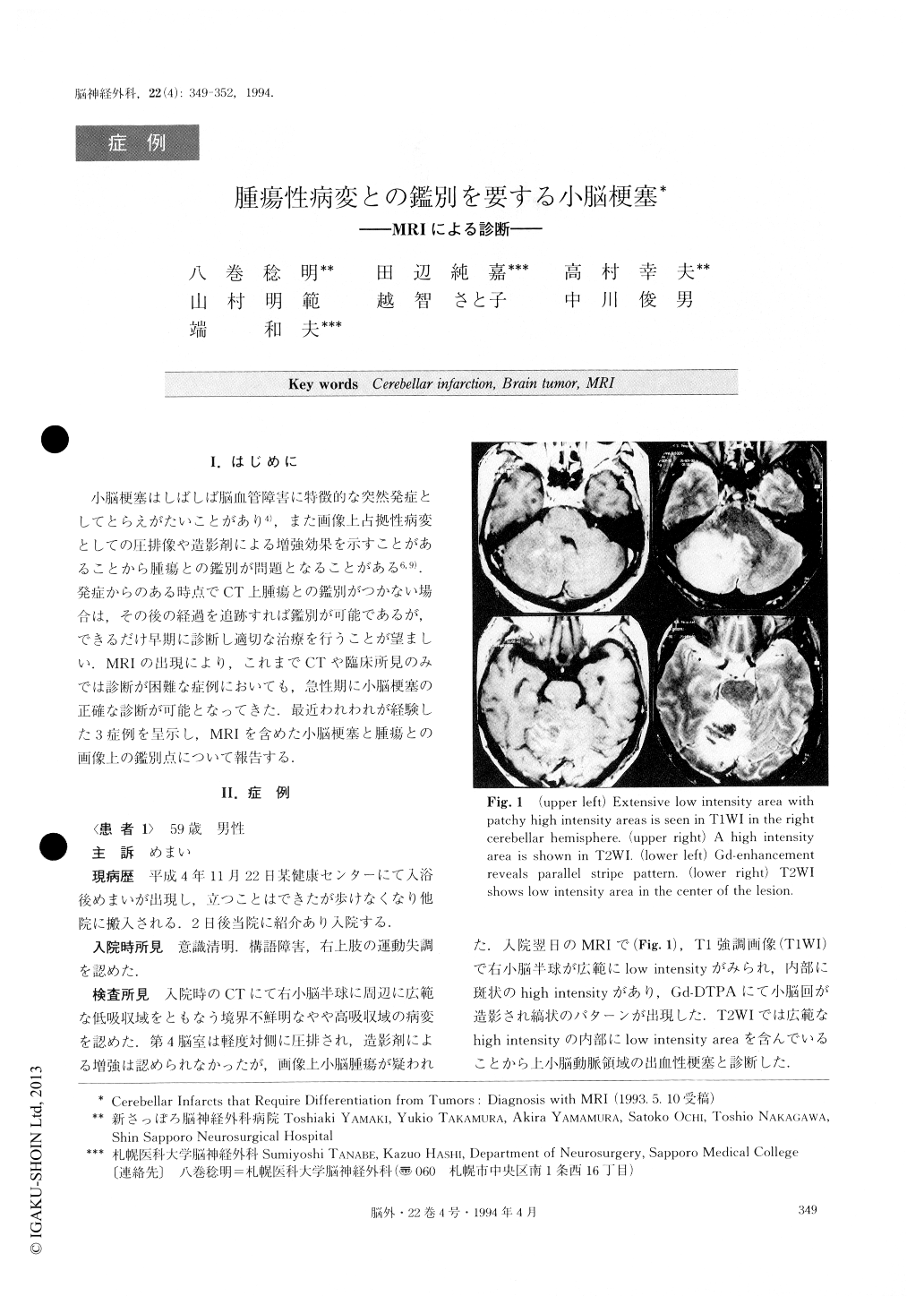
小脳梗塞はしばしば脳血管障害に特徴的な突然発症としてとらえがたいことがあり4),また画像上占拠性病変としての圧排像や造影剤による増強効果を示すことがあることから腫瘍との鑑別が問題となることがある6,9).発症からのある時点でCT上腫瘍との鑑別がつかない場合は,その後の経過を追跡すれば鑑別が可能であるが,できるだけ早期に診断し適切な治療を行うことが望ましい.MRIの出現により,これまでCTや臨床所見のみでは診断が困難な症例においても,急性期に小脳梗塞の正確な診断が可能となってきた.最近われわれが経験した3症例を呈示し,MRIを含めた小脳梗塞と腫瘍との画像上の鑑別点について報告する.
It is often difficult to differentiate cerebellar infarct with cerebellar swelling from neoplastic disorders, be-cause former can be shown as cerebellar mass with marked contrast enhancement on CT scan. We analy-zed radiologically three cases with cerebellar infarction by using MRI, conventional CT scan and angiography.Two cases in the acute stage could be diagnosed as cerebellar infarction by MRI alone based on the follow-ing findings: 1) the lesion was distributed in the terri-tory of cerebellar arteries; 2) the normal pattern of cerebellar folia and fissures was preserved in Gd en-hancement MRI image; 3) characteristics of MRI in-tensity were compatible with hemorrhagic infarction. The other case in the chronic stage showed peculiar en-hancement, which was unusual for infarction. It was dia-gnosed as cerebellar infarction with reference to the angiographic findings. MRI is generally useful to obtain early diagnosis of tumor-like cerebellar infarcts, and proper treatment should be started as early as possible.
Copyright © 1994, Igaku-Shoin Ltd. All rights reserved.