Japanese
English
- 有料閲覧
- Abstract 文献概要
- 1ページ目 Look Inside
I.はじめに
脊髄の解剖学的計測値については今世紀の初めより多くの報告がある.成人では長さ43-45(正常限界40-45)cm,身長の1/4弱で,頸部,腰部に膨大部をもつ.その断面の形状は一定でなく,レベルにより大きく異なる.C6の前後径は9mm,横径は13-14mm,T6の前後径8mm,横径10mm,L3の前後径8.5mm,横径11-13mmなどは臨床解剖上よく知られた数値である12).生体内でこれらの形状,大きさを知ることはCTの出現までおよそ不可能であったが,1970年代末までに椎体,脊椎管のCT解剖はHammerschlagら5),Leeら9),Sheldonら14),Coinら1)により研究され,さらに1980年代にはHarwood-NashとFitz6)は成長期の変化にも注目し,今日ではすでに完成の域に達したと思われる.しかし,脊椎管内の軟部組織はC1-C2のレベルを除いて硬膜外脂肪組織に囲まれた"homogenous density"とみなされ,メトリザマイドCT-myelograplly(以下met.CTと略す)でなければその画像は診断的価値はない2).
met.CTの導入以来,脊髄,神経根糸,馬尾神経,終糸におよばず,前後脊髄動脈までも明らかにされてきている16).
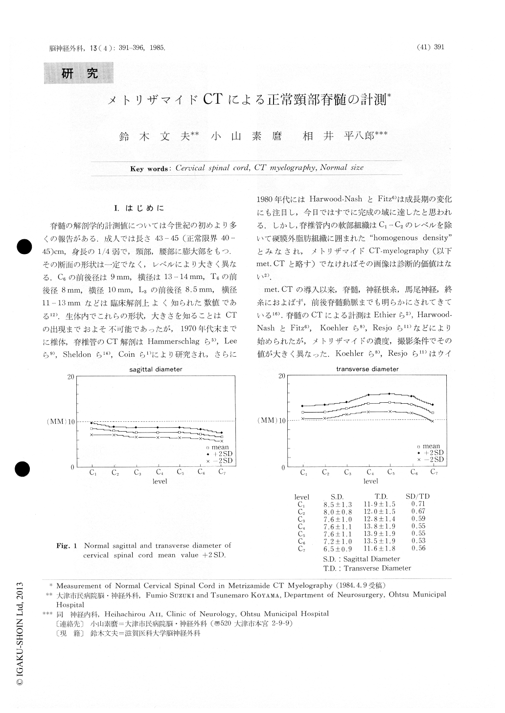
The shape of the spinal cord is the most importantfactor in diagnosis of spinal disorders by metrizamideCT myelography (met. CT). Even in cases wherethe spinal cord looks normal in shape its size mightbe abnormal, for example in cases with spinal cordatrophy, syringomyelia, intramedullary tumor and sev-eral other conditions. In detecting the slightest ab-normality in such cases, it is absolutely necessary tohave in hand the knowledge of the nomal size of thespinal cord at each level. We measured, therefore,the sagittal and transverse diameters of the cervicalspinal cord in 55 patients with no known lesions onmet. CT (Fig. 1).

Copyright © 1985, Igaku-Shoin Ltd. All rights reserved.


