Japanese
English
- 有料閲覧
- Abstract 文献概要
- 1ページ目 Look Inside
- 参考文献 Reference
Ⅰ.はじめに
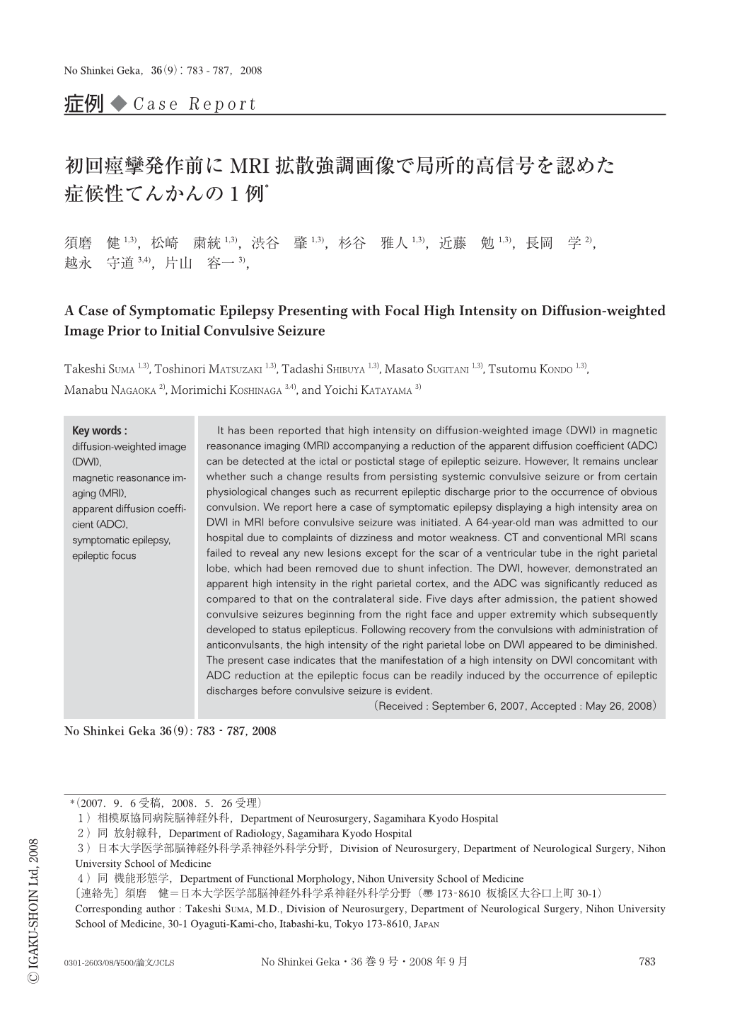
てんかん患者において,痙攣発作直後のCTをはじめとする通常の画像検査では異常所見を認めないことがほとんどである.しかし近年,MRI拡散強調画像(diffusion-weighted image:DWI)において,痙攣発作中ないし発作直後に一過性の高信号が出現することが報告されている2,4,5).今回われわれは,明らかな顔面,四肢の痙攣発作を発症する以前のDWIにおいて,てんかん焦点に一致した高信号の出現を認め,その後に重積発作にまで進行した症候性てんかんの1例を経験したので報告する.
It has been reported that high intensity on diffusion-weighted image (DWI) in magnetic reasonance imaging (MRI) accompanying a reduction of the apparent diffusion coefficient (ADC) can be detected at the ictal or postictal stage of epileptic seizure. However, It remains unclear whether such a change results from persisting systemic convulsive seizure or from certain physiological changes such as recurrent epileptic discharge prior to the occurrence of obvious convulsion. We report here a case of symptomatic epilepsy displaying a high intensity area on DWI in MRI before convulsive seizure was initiated. A 64-year-old man was admitted to our hospital due to complaints of dizziness and motor weakness. CT and conventional MRI scans failed to reveal any new lesions except for the scar of a ventricular tube in the right parietal lobe, which had been removed due to shunt infection. The DWI, however, demonstrated an apparent high intensity in the right parietal cortex, and the ADC was significantly reduced as compared to that on the contralateral side. Five days after admission, the patient showed convulsive seizures beginning from the right face and upper extremity which subsequently developed to status epilepticus. Following recovery from the convulsions with administration of anticonvulsants, the high intensity of the right parietal lobe on DWI appeared to be diminished. The present case indicates that the manifestation of a high intensity on DWI concomitant with ADC reduction at the epileptic focus can be readily induced by the occurrence of epileptic discharges before convulsive seizure is evident.

Copyright © 2008, Igaku-Shoin Ltd. All rights reserved.


