Japanese
English
- 有料閲覧
- Abstract 文献概要
- 1ページ目 Look Inside
Ⅰ.はじめに
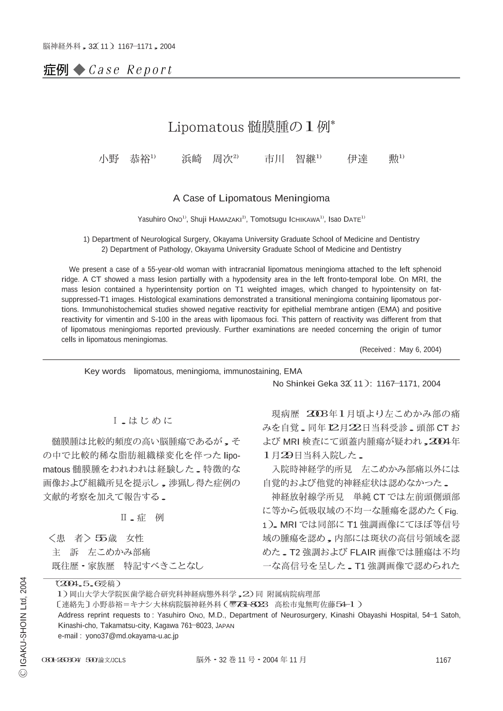
髄膜腫は比較的頻度の高い脳腫瘍であるが,その中で比較的稀な脂肪組織様変化を伴ったlipomatous髄膜腫をわれわれは経験した.特徴的な画像および組織所見を提示し,渉猟し得た症例の文献的考察を加えて報告する.
We present a case of a 55-year-old woman with intracranial lipomatous meningioma attached to the left sphenoid ridge. A CT showed a mass lesion partially with a hypodensity area in the left fronto-temporal lobe. On MRI,the mass lesion contained a hyperintensity portion on T1 weighted images,which changed to hypointensity on fat-suppressed-T1 images. Histological examinations demonstrated a transitional meningioma containing lipomatous portions. Immunohistochemical studies showed negative reactivity for epithelial membrane antigen (EMA) and positive reactivity for vimentin and S-100 in the areas with lipomaous foci. This pattern of reactivity was different from that of lipomatous meningiomas reported previously. Further examinations are needed concerning the origin of tumor cells in lipomatous meningiomas.

Copyright © 2004, Igaku-Shoin Ltd. All rights reserved.


