Japanese
English
解剖を中心とした脳神経手術手技
選択的前床突起除去手技
Selective Anterior Clinoidectomy
糟谷 英俊
1
,
堀 智勝
1
,
馬場 元毅
2
Hidetoshi KASUYA
1
,
Tomokatsu HORI
1
,
Motoki BABA
2
1東京女子医科大学脳神経外科
2東埼玉総合病院脳神経外科
1Department of Neurosurgery,Tokyo Women's Medical University
2Department of Neurosurgery,Higashi-Saitama General Hospital
キーワード:
anterior clinoidectomy
,
anterior clinoid process
,
clinoid space
,
superior orbital fi ssure
,
frontotemporal dural fold (FTDF)
,
meningoorbital band (MOB)
,
optic strut
Keyword:
anterior clinoidectomy
,
anterior clinoid process
,
clinoid space
,
superior orbital fi ssure
,
frontotemporal dural fold (FTDF)
,
meningoorbital band (MOB)
,
optic strut
pp.233-242
発行日 2007年3月10日
Published Date 2007/3/10
DOI https://doi.org/10.11477/mf.1436100426
- 有料閲覧
- Abstract 文献概要
- 1ページ目 Look Inside
- 参考文献 Reference
Ⅰ.はじめに
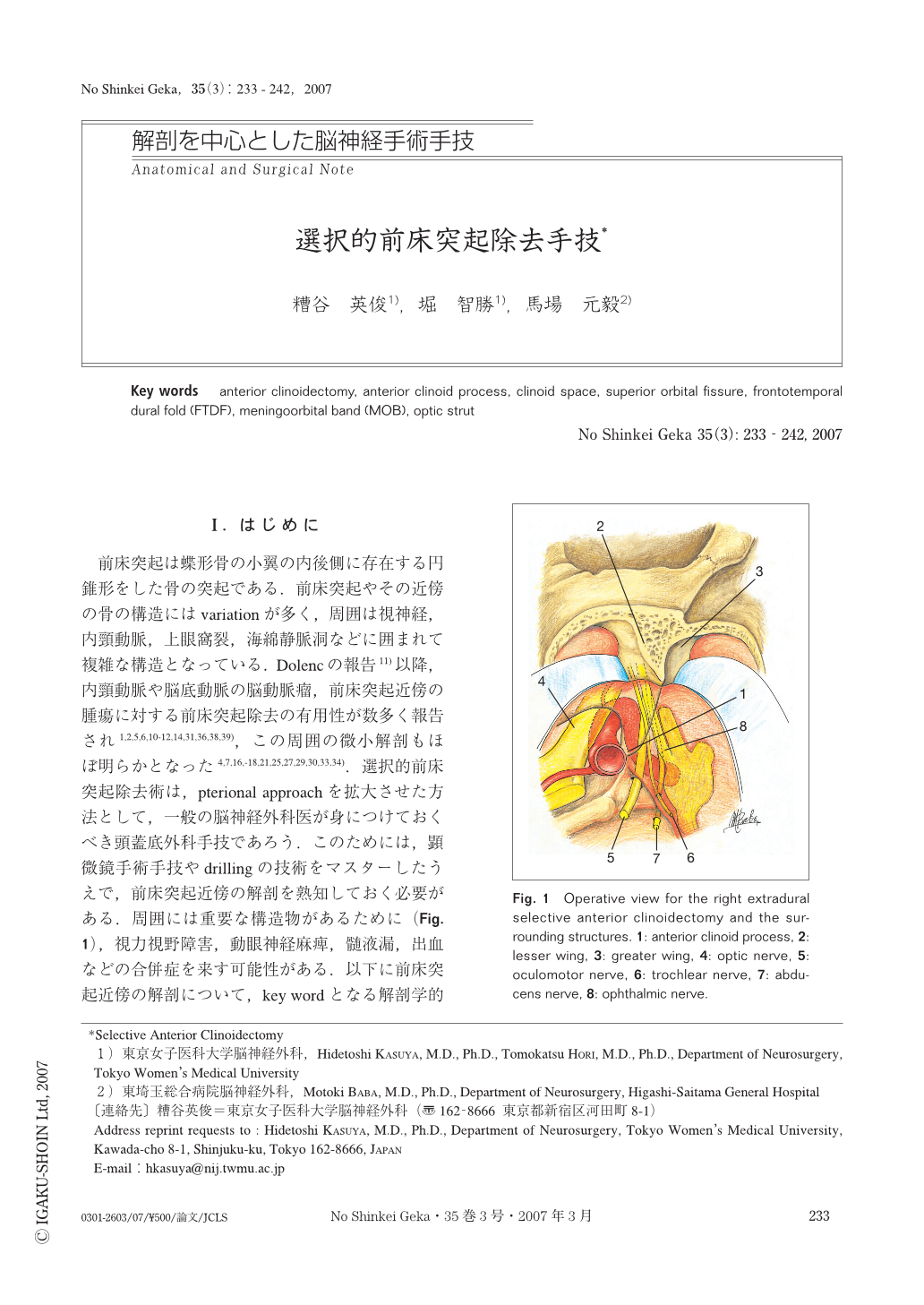
前床突起は蝶形骨の小翼の内後側に存在する円錐形をした骨の突起である.前床突起やその近傍の骨の構造にはvariationが多く,周囲は視神経,内頸動脈,上眼窩裂,海綿静脈洞などに囲まれて複雑な構造となっている.Dolencの報告11)以降,内頸動脈や脳底動脈の脳動脈瘤,前床突起近傍の腫瘍に対する前床突起除去の有用性が数多く報告され1,2,5,6,10-12,14,31,36,38,39),この周囲の微小解剖もほぼ明らかとなった4,7,16,-18,21,25,27,29,30,33,34).選択的前床突起除去術は,pterional approachを拡大させた方法として,一般の脳神経外科医が身につけておくべき頭蓋底外科手技であろう.このためには,顕微鏡手術手技やdrillingの技術をマスターしたうえで,前床突起近傍の解剖を熟知しておく必要がある.周囲には重要な構造物があるために(Fig. 1),視力視野障害,動眼神経麻痺,髄液漏,出血などの合併症を来す可能性がある.以下に前床突起近傍の解剖について,key wordとなる解剖学的用語を説明し,われわれの通常行っている選択的前床突起除去術を解説する.

Copyright © 2007, Igaku-Shoin Ltd. All rights reserved.


