Japanese
English
- 有料閲覧
- Abstract 文献概要
- 1ページ目 Look Inside
- 参考文献 Reference
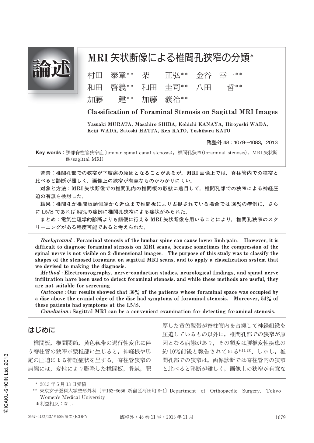
背景:椎間孔部での狭窄が下肢痛の原因となることがあるが,MRI画像上では,脊柱管内での狭窄と比べると診断が難しく,画像上の狭窄が有意なものかわかりにくい.
対象と方法:MRI矢状断像での椎間孔内の椎間板の形態に着目して,椎間孔部での狭窄による神経圧迫の有無を検討した.
結果:椎間孔が椎間板頭側端から近位まで椎間板により占拠されている場合では36%の症例に,さらにL5/Sであれば54%の症例に椎間孔狭窄による症状がみられた.
まとめ:電気生理学的診断よりも簡便に行えるMRI矢状断像を用いることにより,椎間孔狭窄のスクリーニングがある程度可能であると考えられた.
Background:Foraminal stenosis of the lumbar spine can cause lower limb pain. However, it is difficult to diagnose foraminal stenosis on MRI scans, because sometimes the compression of the spinal nerve is not visible on 2-dimensional images. The purpose of this study was to classify the shapes of the stenosed foramina on sagittal MRI scans, and to apply a classification system that we devised to making the diagnosis.
Method:Electromyography, nerve-conduction studies, neurological findings, and spinal nerve infiltration have been used to detect foraminal stenosis, and while these methods are useful, they are not suitable for screening.
Outcome:Our results showed that 36% of the patients whose foraminal space was occupied by a disc above the cranial edge of the disc had symptoms of foraminal stenosis. Moreover, 54% of these patients had symptoms at the L5/S.
Conclusion:Sagittal MRI can be a convenient examination for detecting foraminal stenosis.

Copyright © 2013, Igaku-Shoin Ltd. All rights reserved.


