Japanese
English
- 有料閲覧
- Abstract 文献概要
- 1ページ目 Look Inside
I.はじめに
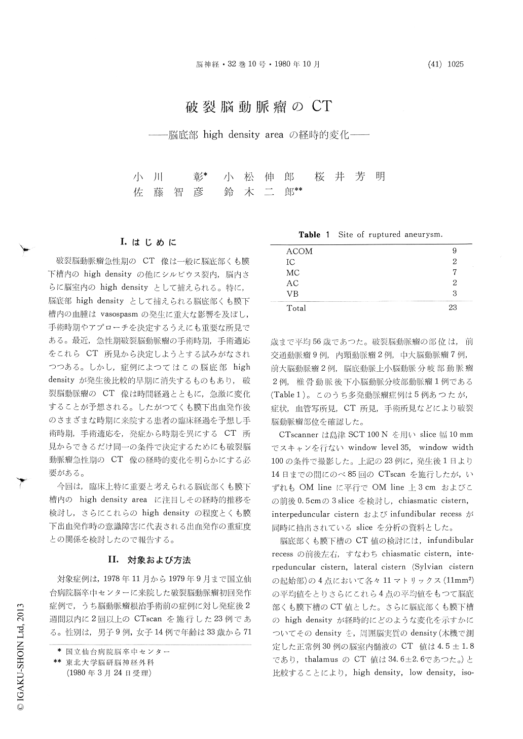
破裂脳動脈瘤急性期のCT像は一般に脳底部くも膜下槽内のhigh densityの他にシルビウス裂内,脳内さらに脳室内のhigh densityとして捕えられる。特に,脳底部high densityとして捕えられる脳底部くも膜下槽内の血腫はvasospasmの発生に重大な影響を及ぼし,手術時期やアプローチを決定するうえにも重要な所見である。最近,急性期破裂脳動脈瘤の手術時期,手術適応をこれらCT所見から決定しようとする試みがなされつつある。しかし,症例によつてはこの脳底部highdensityが発生後比較的早期に消失するものもあり,破裂脳動脈瘤のCT像は時間経過とともに,急激に変化することが予想される。したがつてくも膜下出血発作後のさまざまな時期に来院する患者の臨床経過を予想し手術時期,手術適応を,発症から時期を異にするCT所見からできるだけ同一の条件で決定するためにも破裂脳動脈瘤急性期のCT像の経時的変化を明らかにする必要がある。
今回は,臨床上特に重要と考えられる脳底部くも膜下槽内の high density area に注目しその経時的推移を検討し,さらにこれらのhigh densityの程度とくも膜下出血発作時の意識障害に代表される出血発作の重症度との関係を検討したので報告する。
Using the findings of CT scanning in cases ofsubarachnoid hemorrhage, we have undertaken an investigation of the relationship between the degree of consciousness disturbance at the time of sub-arachnoid hemorrhage and the degree of high density area within the basal cistern within the 1st day from onset, and the sequential changes of the high density areas.
The study included 23 cases of initial attacks of cerebral aneurysm rupture in which 2 or more CT scans were obtained prior to surgery, or in non-operated cases, within 2 weeks of onset.
The severity of the attacks were classified into 3 groups: minor attack, cases of subarachnoid hemorrhage without loss of the consciousness ; moderate attack, those with loss of consciousness extending for less than 1 hour; and major attack, those with loss of consciousness extending for more than 1 hour.
Since a correlation was found between the CT number of high density areas in the basal cistern within the first 24 hours from onset and the severity of the attack, the classification of the severity of the attack is thought to correlate with the volume of the subarachnoid hemorrhage.
Disappearance of the high density areas in the basal cistern occurred after about 2 days in the minor, about 4 days in the moderate, and 8-10 days in the major attack cases, but in the group of patients suffering recurrent attacks and in which death occurred without a return of consciousness, the high density areas remained until death.
We conclude that by considering the CT number of the high density areas obtained by CT scanning in the basal cistern in cases of subarachnoid hemor-rhage, the degree of bleeding can be determined, and the incidence of infarction due to vasospasm can be anticipated.

Copyright © 1980, Igaku-Shoin Ltd. All rights reserved.


