Japanese
English
- 有料閲覧
- Abstract 文献概要
- 1ページ目 Look Inside
I.はじめに
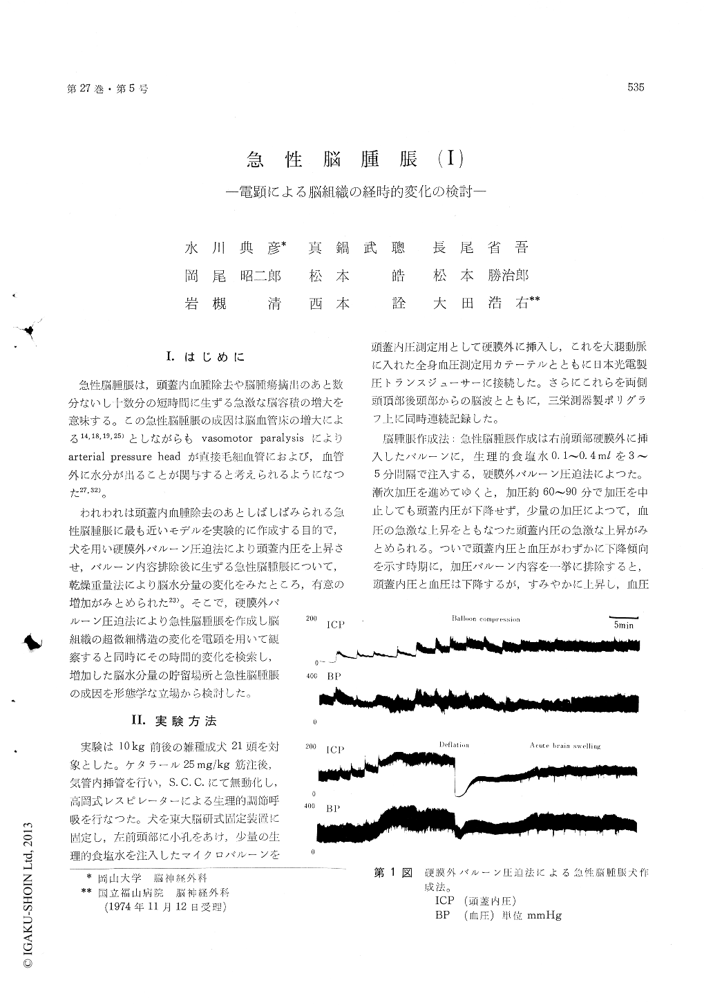
急性脳腫脹は,頭蓋内血腫除去や脳腫瘍摘出のあと数分ないし十数分の短時間に生ずる急激な脳容積の増大を意味する。この急性脳腫脹の成因は脳血管床の増大による14,18,19,25)としながらもvasomotor paralysisによりarterial pressure headが直接毛細血管におよび,血管外に水分が出ることが関与すると考えられるようになつた27,32)。
われわれは頭蓋内血腫除去のあとしばしばみられる急性脳腫脹に最も近いモデルを実験的に作成する目的で,犬を用い硬膜外バルーン圧迫法により頭蓋内圧を上昇させ,バルーン内容排除後に生ずる急性脳腫脹について,乾燥重量法により脳水分量の変化をみたところ,有意の増加がみとめられた23)。そこで,硬膜外バルーン圧迫法により急性脳腫脹を作成し脳組織の超微細構造の変化を電顕を用いて観察すると同時にその時間的変化を検索し,増加した脳水分量の貯留場所と急性脳腫脹の成因を形態学な立場から検討した。
We used 21 adult dogs and produced acute brainswelling by extradural balloon compression method.Left parietal brain tissues were examined electron-microscapically at immediately (5 dogs), 30 min.(8 dogs) and one or two hours (5 dogs) after theonset of the rebound phenomenon following balloondeflation.
The findings were as follows:
1) Immediately after the rebound phenomenonfollowing balloon deflation, pericapillary astroglialswelling and dilated capillary vessels were morefrequently found than control, but no edematouschanges in neuropil nor nerve cell changes wereobserved.
2) 30 min. after the rebound phenomenon, peri-capillary astroglial swelling became more markedand edematous changes in neuropil were detected.The mitochondrial swelling and resolution of cristaein neuron became apparent and about one half ofthe nerve cells showed degenerative changes. Astro-glial swelling and sometimes presynapic or dendriticswelling were observed.
3) One hour or 2 hrs. after the rebound phe-nomenon, almost all of the nerve cells showed de-generative changes. The mitochondrial changes inneuron became marked but no apparent extracellulardilatation in the gray and white matter were de-tected. The endothelial swelling bacame obvious.
4) Throughout the course, the ultrastructuralchanges in the white matter were not so markedas in the gray matter. Slightly distended extra-cellular space and resolution of myeline sheath ordilatation of periaxonal space were detected. Theseresults led us to the conclusions:
a) In an early phase, acute brain swelling wasconsidered to be due to enlarged cerebrovascularbed and pericapillary astroglial swelling.
b) About 30 min. after the rebound phenomenonfollowing balloon deflation, the edematous factorin acute brain swelling became apparent and moreprominent one hour or 2 hrs. after the rebound.
c) Nerve cell changes with mitochondrial swell-ing and resolution of the cristae were detectedThese findings began to appear 30 min. after therebound, and almost all of the neuron showed thesefindings one or 2 hrs. after the rebound. Thesechanges were probably due to the brain ischemiacaused by impaired CBF.
d) Throughout the course, there was no markedexpansion of extracellular space in the white matter.
Copyright © 1975, Igaku-Shoin Ltd. All rights reserved.