Japanese
English
- 有料閲覧
- Abstract 文献概要
- 1ページ目 Look Inside
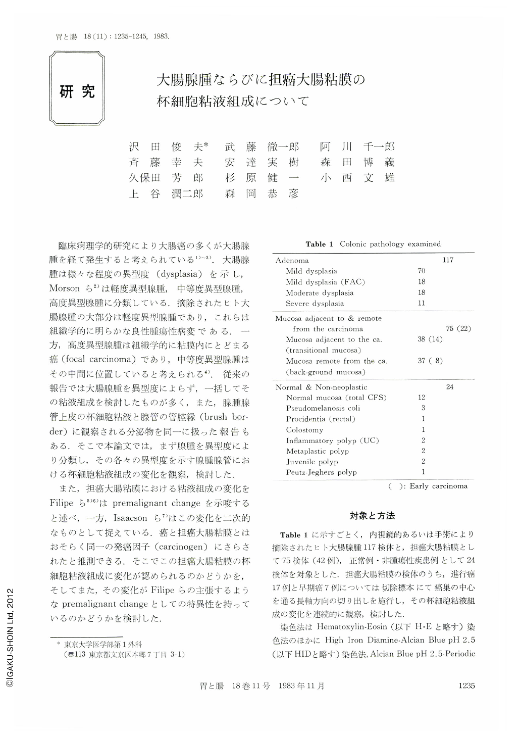
臨床病理学的研究により大腸癌の多くが大腸腺腫を経て発生すると考えられている1)~3).大腸腺腫は様々な程度の異型度(dysplasia)を示し,Morsonら2)は軽度異型腺腫,中等度異型腺腫,高度異型腺腫に分類している.摘除されたヒト大腸腺腫の大部分は軽度異型腺腫であり,これらは組織学的に明らかな良性腫瘍性病変である.一方,高度異型腺腫は組織学的に粘膜内にとどまる癌(focal carcinoma)であり,中等度異型腺腫はその中間に位置していると考えられる4).従来の報告では大腸腺腫を異型度によらず,一括してその粘液組成を検討したものが多く,また,腺腫腺管上皮の杯細胞粘液と腺管の管腔縁(brush border)に観察される分泌物を同一に扱った報告もある.そこで本論文では,まず腺腫を異型度により分類し,その各々の異型度を示す腺腫腺管における杯細胞粘液組成の変化を観察,検討した.
また,担癌大腸粘膜における粘液組成の変化をFilipeら5)6)はpremalignant changeを示唆すると述べ,一方,Isaacsonら7)はこの変化を二次的なものとして捉えている.癌と担癌大腸粘膜とはおそらく同一の発癌因子(carcinogen)にさらされたと推測できる.そこでこの担癌大腸粘膜の杯細胞粘液組成に変化が認められるのかどうかを,そしてまた,その変化がFilipeらの主張するようなpremalignant changeとしての特異性を持っているのかどうかを検討した.
One hundred and seventeen adenomas and 42 cases of resected specimens of carcinomas of the large intestine were investigated by H-E, HID-AB (High Iron Diamine-Alcian Blue, pH 2.5), AB (pH 2.5)-PAS and PKP (PAT/KOH/PAS: Periodic Acid Thionine Schiff/Potassium Hydroxide/Periodic Acid Schiff) stain in order to see mucin characteristics of these lesions. As a control, 24 sections of normal and non-neoplastic mucosa were also examined.
Sixty-six percent of adenomas with mild dysplasia showed increased sulphomucin (brown), whereas mucin components of adenomas in familial polyposis coli (FAC) with mild dysplasia and adenomas with moderate and severe dysplasia showed increase of sialomucin (blue) (56%, 61%, 54%) in HID-AB stain. The higher the dysplasia, the more sialomucin appeared. The transitional mucosa showed characteristic increase of sialomucin (76%) and the background mucosa also represented increased components of sialomucin in both oral and anal sites (61%). However, increase of sialomucin was also found in normal mucosa and also in mucosa of non-neoplastic disease such as rectal procidentia, colostomy, pseudomelanosis coli. In AB-PAS stain, acid mucin (blue) was predominant in adenomas of FAC wich mild dysplasia and adenomas with moderate and severe dysplasia (56%, 61%, 45%), and also in the transitional mucosa of advanced carcinomas (67%). In PKP stain 70% of adenomas with mild dysplasia showed C8-O-acetylated or diacetylated sialomucin, whereas 60% of adenomas with moderate and severe dysplasia showed coexistance of non-acetylated or C7- or C9-O-acetylated sialomucin. On the other hand, 58% of sialomucin of transitional and background mucosa were C8-O-acetylated or diacetylated in PKP stain.
From our observations, it is clear that the transitional mucosa, background mucosa and adenomas with moderate dysplasia showed increase of sialomucin in HID‐AB stain. However, as increase of sialomucin was also observed in normal and non-neoplastic disease, the mucosa showing predominant sialomucin was not assumed to represent malignant transformation in the large intestine. In addition to these facts, PKP stain revealed that O‐acetylated sialomucin was predominant in the transitional and background mucosa, whereas non-acetylated sialomucin was predominaret in adenomas with moderate or severe dysplasia. Although these non-acetylated sialomucin was observed in the normal colonic mucosa, it is unclear whether these non-acetylated sialomucin in adenomas with moderate or severe dysplasia has the same characteristics of those in normal colonic mucosa. Further detailed study is required to clarify the fine characteristics of goblet cell mucins in the transitional mucosa, adenomas, carcinomas and non-neoplastic diseases.

Copyright © 1983, Igaku-Shoin Ltd. All rights reserved.


