Japanese
English
- 有料閲覧
- Abstract 文献概要
- 1ページ目 Look Inside
- サイト内被引用 Cited by
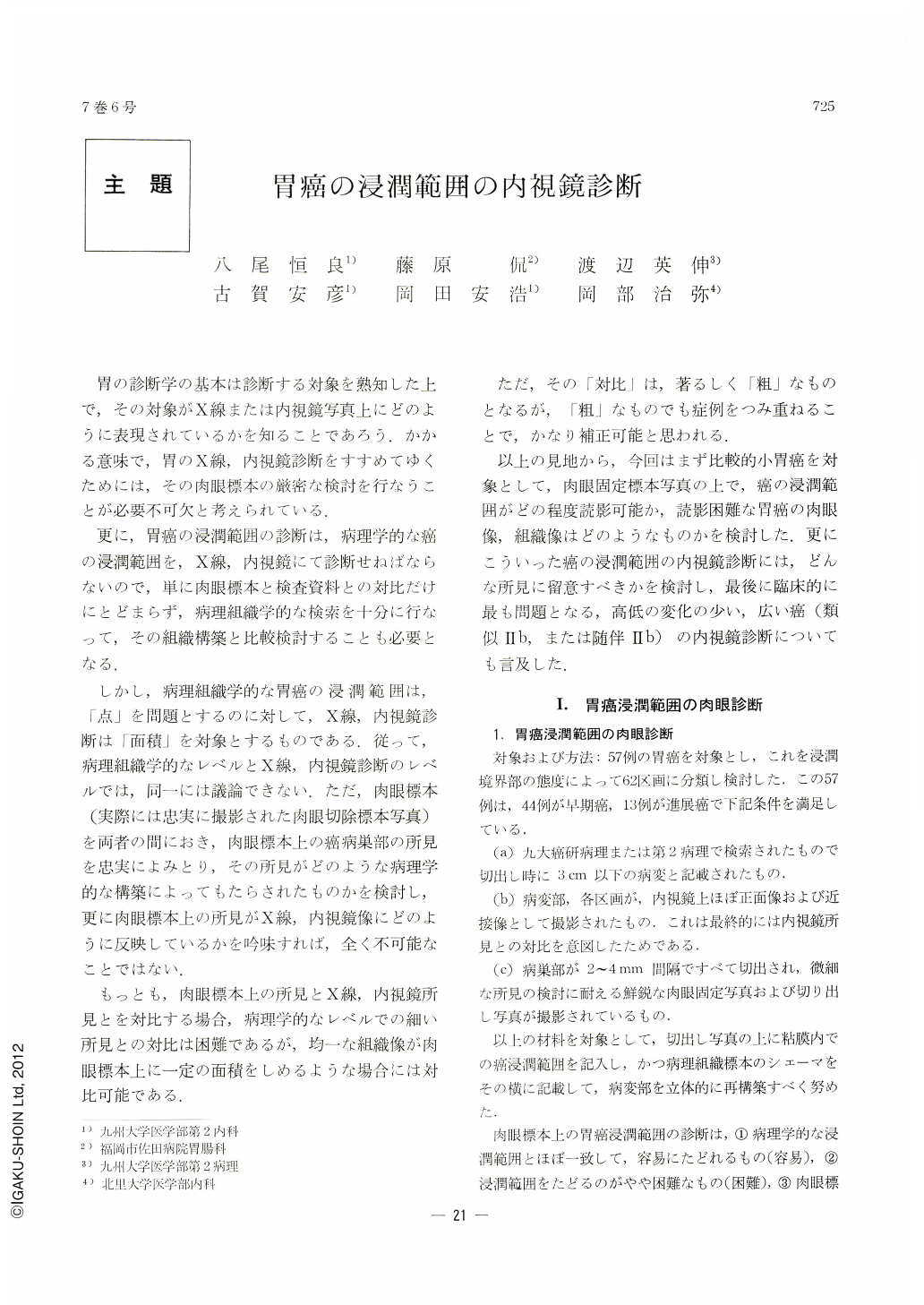
胃の診断学の基本は診断する対象を熟知した上で,その対象がX線または内視鏡写真上にどのように表現されているかを知ることであろう.かかる意味で,胃のX線,内視鏡診断をすすめてゆくためには,その肉眼標本の厳密な検討を行なうことが必要不可欠と考えられている.
更に,胃癌の浸潤範囲の診断は,病理学的な癌の浸潤範囲を,X線,内視鏡にて診断せねばならないので,単に肉眼標本と検査資料との対比だけにとどまらず,病理組織学的な検索を十分に行なって,その組織構築と比較検討することも必要となる.
In order to know the limitations of endoscopic diagnosis in estimating the extent of infiltration of gastric cancer, we have studied 57 cases of relatively small cancers. To evaluate their gross findings, histological structures and capability of endoscopic diagnosis, we divided them into 62 small areas.
1) Gastric cancer most hard to diagnose belonged to that variety showing what Fujiwara calls superficial type. The surface of the mucosa in this variety was covered with non-cancerous epithelia and the lesions showed area-like pictures.
2) When stratum of cancer nests was thin and seen only in the intermediate or deeper parts of the mucosal layer, its diagnosis was impossible not only macroscopically but also even by endoscopy.
3) Undifferentiated carcinoma was apt to been seen as discoloration of the affected part, while well differentiated carcinoma tended to be confirmed by its engorgement.
4) Endoscopic diagnosis of cancer mentioned in (1) necessitated careful reading not only of minute shades of difference in mucosal hue but also of detailed patterns such as transparent small blood vessels. The cancer in question was characterized by gradual discoloration of the mucosal surface with its borders indistinct and also by changes of minute patterns on it. Discoloration began from within the lesion and extended outward.
5) When orderly minute patterns were observed on the mucosal surface, no exposed cancer cancer existed over it.
We have also investigated 11 cases of those small cancers whose surface was almost of the same height as the surrounding mucosa, and following results were obtained.
1) Endoscopic estimation of cancer borders depends more on the gross pictures and histological structures than on their difference in height.
2) Some of highly differentiated adenocarcinoma can be diagnosed with ease, even though estimation of its borders be impossible.

Copyright © 1972, Igaku-Shoin Ltd. All rights reserved.


