Japanese
English
- 有料閲覧
- Abstract 文献概要
- 1ページ目 Look Inside
患 者:46歳 男.
主 訴:黄疸.
昭和46年10月悪心吐嘔,12月末高度黄疸,一時軽減するも再度黄疸増強,47年6月当科受診,第2外科へ入院
現 症:黄疸著明,心窩部3横指肝触知.
Case : A 68 years old man, 6 months before admission, complainning of jaundice and itching without abdominal pain, visited our clinic. Both family and past histories were not noteworthy.
Laboratory examination revealed that the liver function was slightly disturbed, and post hepatic obstruction was suspected. Pancreozimin-Secretin-test showed prominent pancreatic exocrine disturbance.
Liver scintiscanning test was normal.
Usual stomach examination was not remarkable.
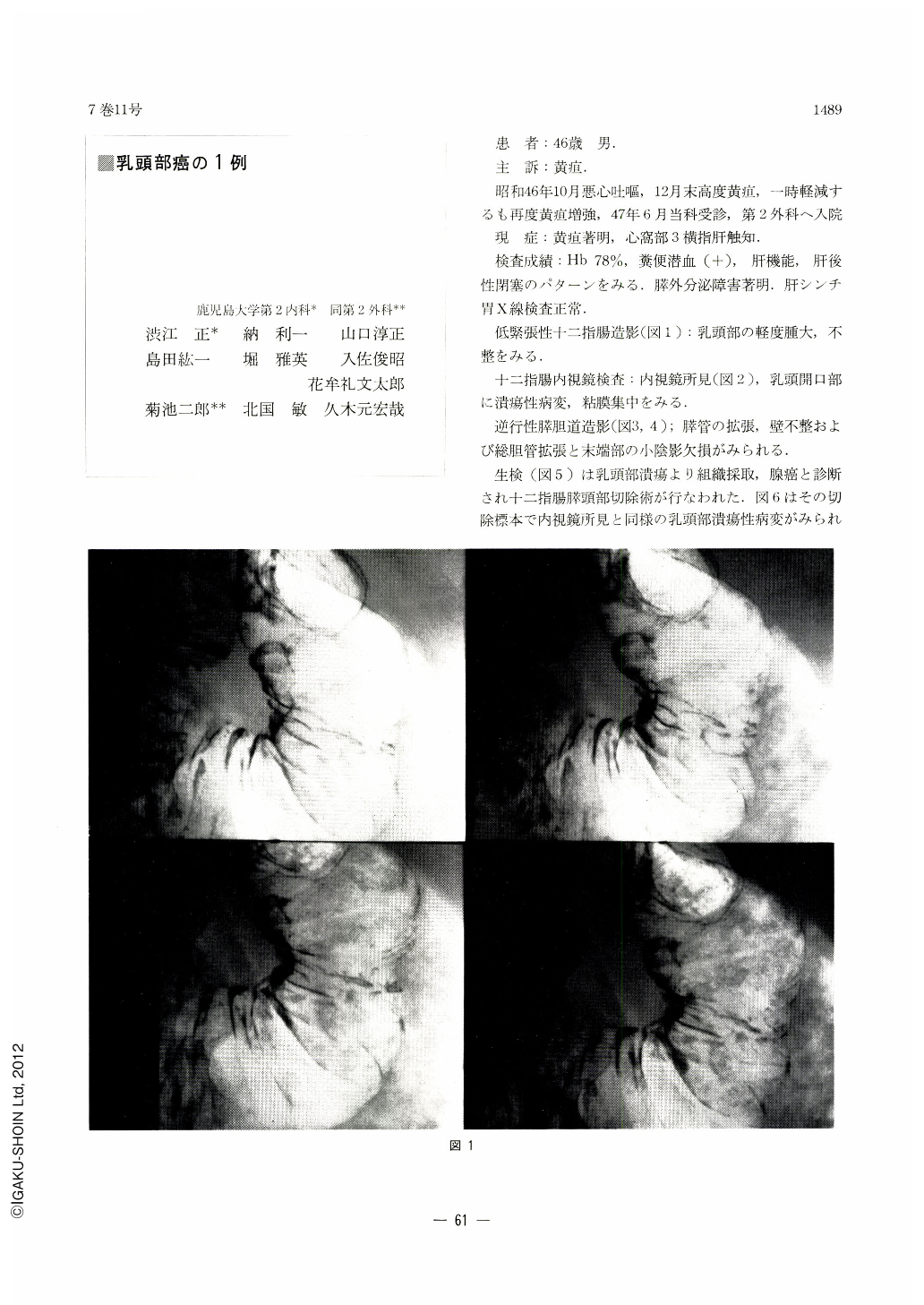
Hypotonic duodenography (Fig. 1) revealed that the Vater papilla was swollen and irregular in shape.
Fiberduodenoscopic examination revealed that the orifice of the Vater papilla was ulcerated and convergence of the folds to it was observed (Fig. 2).
Endoscopic pancreatochol angiography (Fig. 3, 4) revealed dilated main pancreatic duct with irregularity of the caliber.
The common bile duct was dilated and the defect of shadow was seen in the region of the terminal choledocus.
Histological examination of biopsy specimens revealed adenocarcinoma involving the duodenal mucosa at Vater's papilla (Fig. 5).
Macroscopic findings.
Resected specimen showed ulcerated orifice of the Vater papilla and convergance of the fold to it.
Cut surface of the tumor mass measured 15×7 mm in diameter (Fig. 6).
Microscopic findings.
There was well differentiated adenocarcinoma in duodenal papilla region with minimal invasion into the head of the pancreas. The tumor appears to originate in either the pancreas duct or common bile duct epithelium at the papilla region (Fig. 7).

Copyright © 1972, Igaku-Shoin Ltd. All rights reserved.


