Japanese
English
- 有料閲覧
- Abstract 文献概要
- 1ページ目 Look Inside
- サイト内被引用 Cited by
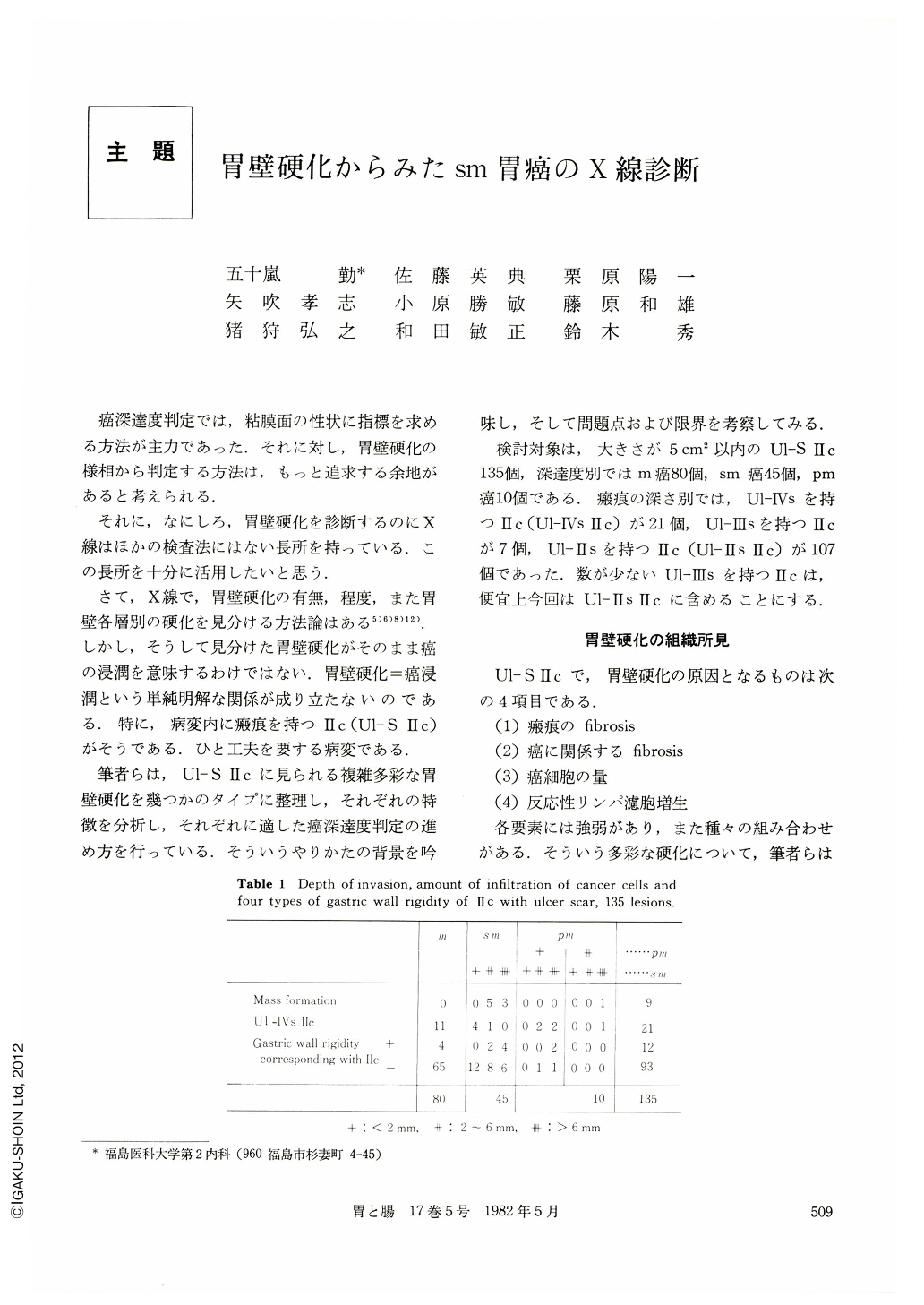
癌深達度判定では,粘膜面の性状に指標を求める方法が主力であった.それに対し,胃壁硬化の様相から判定する方法は,もっと追求する余地があると考えられる.
それに,なにしろ,胃壁硬化を診断するのにX線はほかの検査法にはない長所を持っている.この長所を十分に活用したいと思う.
It is troublesome to detect the depth of invasion of the cancer in Ⅱc type lesion with ulcer scars. This is because it has many various types of gastric wall rigidity.
The authors have categorized radiologically these variegated gastric wall rigidity into four types based on the presence of mass formation of the submucosa and rigidity of the whole lesion. Moreover, characteristics of each type were analyzed (Table 1), and the method of judging the depth of invasion suitable to each type was shown in Table 2.
1. Ⅱc with mass formation in the submucosa
Depth of invasion was more than 2 to 6 mm in the submucosa and not in the mucosa. The mass formation could be detectable by compression study, however, it was difficult to reveal by the macroscopic observations of the mucosa.
2. Ⅱc with Ul-Ⅲs
Determining the depth of invasion was impossible. Most of the lesions were located in the gastric angle.
3. Other than the above-mentioned two types, Ⅱc with gastric wall rigidity over the entire lesion was observed. In cases located in regions other than the gastric angle, cancer cells invade either the submucosa (++) or proper muscle layer. In the other-cases located in the gastric angle, the cancer cells invaded the mucosa.
4. Ⅱc other than the three types mentioned
Gastric wall rigidity was seen in parts around the ulcer scar. In this type of Ⅱc, cancer cells invaded the submucosa (++~+++). In cases showing rigidity in the region of the ulcer scar only, cancer cells invaded either mucosa or the submucosa (+).

Copyright © 1982, Igaku-Shoin Ltd. All rights reserved.


