Japanese
English
- 有料閲覧
- Abstract 文献概要
- 1ページ目 Look Inside
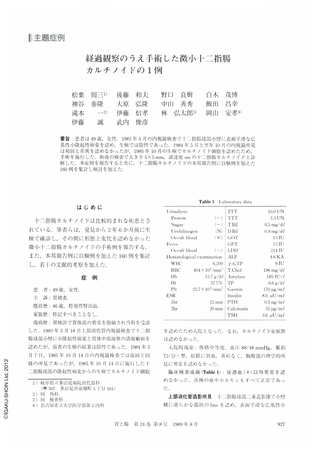
要旨 患者は49歳,女性.1983年5月の内視鏡検査で十二指腸球部小彎に表面平滑な広基性小隆起性病変を認め,生検では陰性であった.1984年5月と翌年10月の内視鏡所見は初回と差異を認めなかったが,1985年10月の生検でカルチノイド細胞を認めたため,手術を施行した.術後の検索で大きさ5×5mm,深達度smの十二指腸カルチノイドと診断した.本症例を報告すると共に,十二指腸カルチノイドの本邦報告例に自験例を加えた160例を集計し検討を加えた.
A rare case of minute carcinoid tumor in the duodenal bulb is reported. A 49-year-old woman visited our hospital requesting a further examination of the stomach because of a lesion in the corpus noted at another hospital. On barium x-ray and endoscopy, although the corpus lesion proved to be a benign ulcer scar, a small polypoid lesion was found on the lesser curvature of the duodenal bulb. Covered with normal mucosa, the lesion was slightly depressed at its top with a reddish appearance. Biopsy from the top failed to lead to a histologic diagnosis. No significant changes in the lesion occurred both in size and gross appearance on followup endoscopy performed twice, at 1 and 2.5 years after the first examination. However, one of the biopsy specimens obtained at the last endoscopy consisted of histological structures compatible with carcinoid tumor. Gastroduodenotomy was undertaken 1.5 months after the last biopsy. The tumor was 5×5 mm in size and localized in the submucosal layer. Immunohistochemical examination showed that the tumor was truly a carcinoid comprising EGC (endocrine granule constituent) reactive cells with positive reaction to argyrophil, but not to argentaffin, cells.

Copyright © 1989, Igaku-Shoin Ltd. All rights reserved.


