Japanese
English
- 有料閲覧
- Abstract 文献概要
- 1ページ目 Look Inside
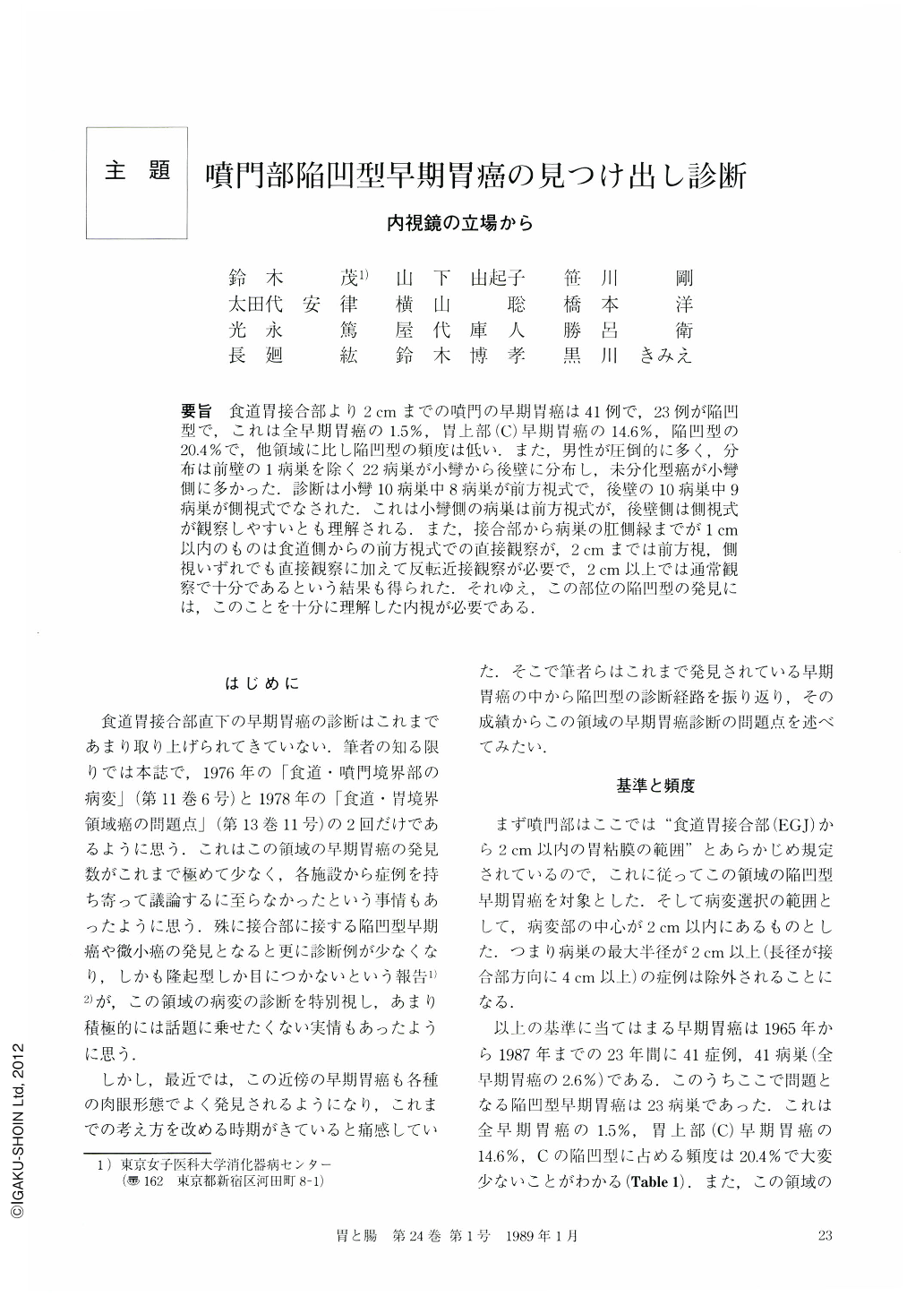
要旨 食道胃接合部より2cmまでの噴門の早期胃癌は41例で,23例が陥凹型で,これは全早期胃癌の15%,胃上部(C)早期胃癌の14.6%,陥凹型の20.4%で,他領域に比し陥凹型の頻度は低い.また,男性が圧倒的に多く,分布は前壁の1病巣を除く22病巣が小彎から後壁に分布し,未分化型癌が小彎側に多かった.診断は小彎10病巣中8病巣が前方視式で,後壁の10病巣中9病巣が側視式でなされた.これは小彎側の病巣は前方視式が,後壁側は側視式が観察しやすいとも理解される.また,接合部から病巣の肛側縁までが1cm以内のものは食道側からの前方視式での直接観察が,2cmまでは前方視,側視いずれでも直接観察に加えて反転近接観察が必要で,2cm以上では通常観察で十分であるという結果も得られた.それゆえ,この部位の陥凹型の発見には,このことを十分に理解した内視が必要である.
Endoscopic diagnosis of depressed type of early gastric cancer located nearby the esophagogastric junction (EGJ) has been considered to be very difficult because of anatomical and functional reasons associated with this region. Therefore, endoscopic diagnosis of the depressed type in the cardia within 2 cm of the EGJ was studied in this paper.
Forty one cases of early gastric cancer have been detected endoscopically in this region during a period of 23 years from 1965 to 1987, and 23 cases of 41 have been of depressed type. The incidence of these was 1.5% of the total number of cases of early gastric cancer, 14.6% of all cases of early gastric cancer in the upper part of the stomach (C), and 20.4% of all cases of depressed type of C. This incidence is very low in comparison with that incidence of depressed type in other regions. Nineteen cases (82.6%) were males, and 22 cases were distributed from the lesser curvature to the posterior wall.
The first endoscopic detection in 8 of 10 cases of depressed type located on the lesser curvature was made with the forward viewing type of fiberscope, and in 9 of 10 cases of depressed types located on the posterior wall detection was made with the lateral viewing type. This result indicates that a depressed lesion on the lesser curvature might be easily detected with the forward viewing type, and a depressed lesion on the posterior wall might be better detected with the lateral viewing type. Furthermore, the distance from EGJ to the anal margin of the depressed lesion was very important for the selection of the types of the scopes and endoscopic biopsy. In other words, a lesion located within 1 cm of EGJ must be directly observed from the esophageal side with the forward viewing type. A lesion within 1~2 cm of EGJ must be observed by direct observation and by the U-turn method with a forward or lateral viewing type. A lesion located beyond 2 cm might be easily observed by routine techniques with both types. Therefore, in order to detect more lesions of the depressed type, the facts mentioned above must be correctly understood for endoscopy of this region.

Copyright © 1989, Igaku-Shoin Ltd. All rights reserved.


