Japanese
English
- 有料閲覧
- Abstract 文献概要
- 1ページ目 Look Inside
- サイト内被引用 Cited by
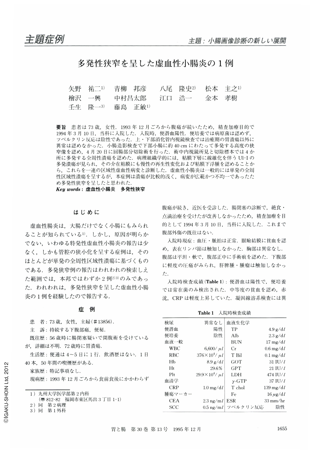
要旨 患者は73歳,女性.1993年12月ごろから腹痛が続いたため,精査加療目的で1994年3月10日,当科に入院した.入院時,便潜血陽性.便培養では病原菌は認めず,ツベルクリン反応は陰性であった.上・下部消化管内視鏡検査では治癒期の胃潰瘍以外に異常は認めなかった.小腸造影検査で下部小腸に約40cmにわたって多発する高度の狭窄像を認め,4月20日に回腸部分切除術を行った.術中内視鏡所見と切除標本では4か所に多発する全周性潰瘍を認めた.病理組織学的には,粘膜下層に線維化を伴うUl-Iの多発潰瘍が見られ,その介在粘膜にも慢性の再生性変化および粘膜下浮腫を認めることから,これらを一連の区域性虚血性病変と診断した.虚血性小腸炎は一般的には単発の全周性区域性潰瘍を呈するが,本症例は潰瘍が比較的浅く,病変が広範かつ不均一であったため多発性狭窄を呈したと思われた.
A 73-year-old woman was admitted with a complaint of lower abdominal pain which had lasted for three months. She had no history of abdominal trauma. On admission, feces was positive for occult blood and negative for pathogenic bacterium. Esophagogastroduodenoscopy and colonoscopy were unremarkable except for a healing ulcer of the stomach. The abdominal ultrasonography demonstrated extensive wallthickening of the ileum. The radiographic examination of the small bowel showed multiple strictures of the distal ileum measuring about 40 cm in length. Resected specimen of the diseased ileum demonstrated four circumferential shallow ulcerations. Histologically, multiple ulcerations were accompanied by submucosal fibrosis and the surrounding areas revealed regenerative mucosal changes due to chronic inflammation and edematous submucosa. Therefore, all these lesions were considered to be parts of a segmental Ischemic lesion.

Copyright © 1995, Igaku-Shoin Ltd. All rights reserved.


