Japanese
English
- 有料閲覧
- Abstract 文献概要
- 1ページ目 Look Inside
- サイト内被引用 Cited by
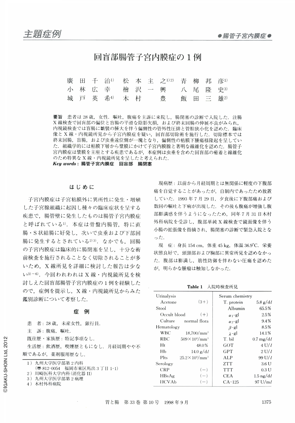
要旨 患者は28歳,女性.嘔吐,腹痛を主訴に来院し,腸閉塞の診断で入院した.注腸X線検査で回盲部の偏位と盲腸の平滑な陰影欠損,および終末回腸の伸展不良がみられ,内視鏡検査では盲腸に皺襞の腫大を伴う偏側性の管外性圧排と管腔狭小化を認めた.臨床像とX線・内視鏡所見から子宮内膜症を疑い,回盲部切除術を施行した.切除標本では終末回腸,盲腸,および虫垂遠位側が一塊となり,偏側性の粘膜下腫瘍様隆起を呈していた.組織学的には粘膜下層から漿膜にかけて子宮内膜腺と著明な線維化を認めた.腸管子宮内膜症は漿膜を主座とする疾患であるが,本症例は虫垂を含めた回盲部の癒着と線維化のため特異なX線・内視鏡所見を呈したと考えられた.
A 28-year-old female was admitted in July, 1993, under the diagnosis of intestinal obstruction. Barium enema examination revealed that the patient had a smooth and extrinsic filling defect at the ileocecal valve and stricturing in the terminal ileum. Colonoscopy also revealed an eccentric submucosal tumor in the cecum, from which normal colonic mucosa was obtained with biopsy forceps, and stricturing of the ileocecal valve. The patient was treated by ileocecal resection under the tentative diagnosis of intestinal endometriosis. Histological examination of the resected specimen revealed that the adhesed ileum, cecum, and appendix was composed of nonneoplastic endometrial tissue infiltrating into the submucosa, the proper muscular layer and the serosa, and there was marked fibrosis in the latter two layers. These findings suggest that submucosal protrusion accompanied by stricturing is one of the features characteristic of intestinal endometriosis.

Copyright © 1998, Igaku-Shoin Ltd. All rights reserved.


