Japanese
English
- 有料閲覧
- Abstract 文献概要
- 1ページ目 Look Inside
I.はじめに
非外傷性頭蓋内解離性動脈瘤(DA)は椎骨脳底動脈系に多く,内頸動脈系に少ないと報告されている16).一般に,DAの診断は脳血管撮影における特徴的な所見1,8),MRI検査での壁内血腫の同定5)や剖検により行われている.内頸動脈系DAは脳梗塞症状で発症し16),脳血管撮影所見も狭窄や閉塞を示す症例が多いので,その診断に苦慮することがある.最近,われわれは2例の中大脳動脈DAを経験したので,症例を提示してその診断や治療における問題点を検討し報告する.また,いままでの報告例をreviewしてその特徴を明らかにする.
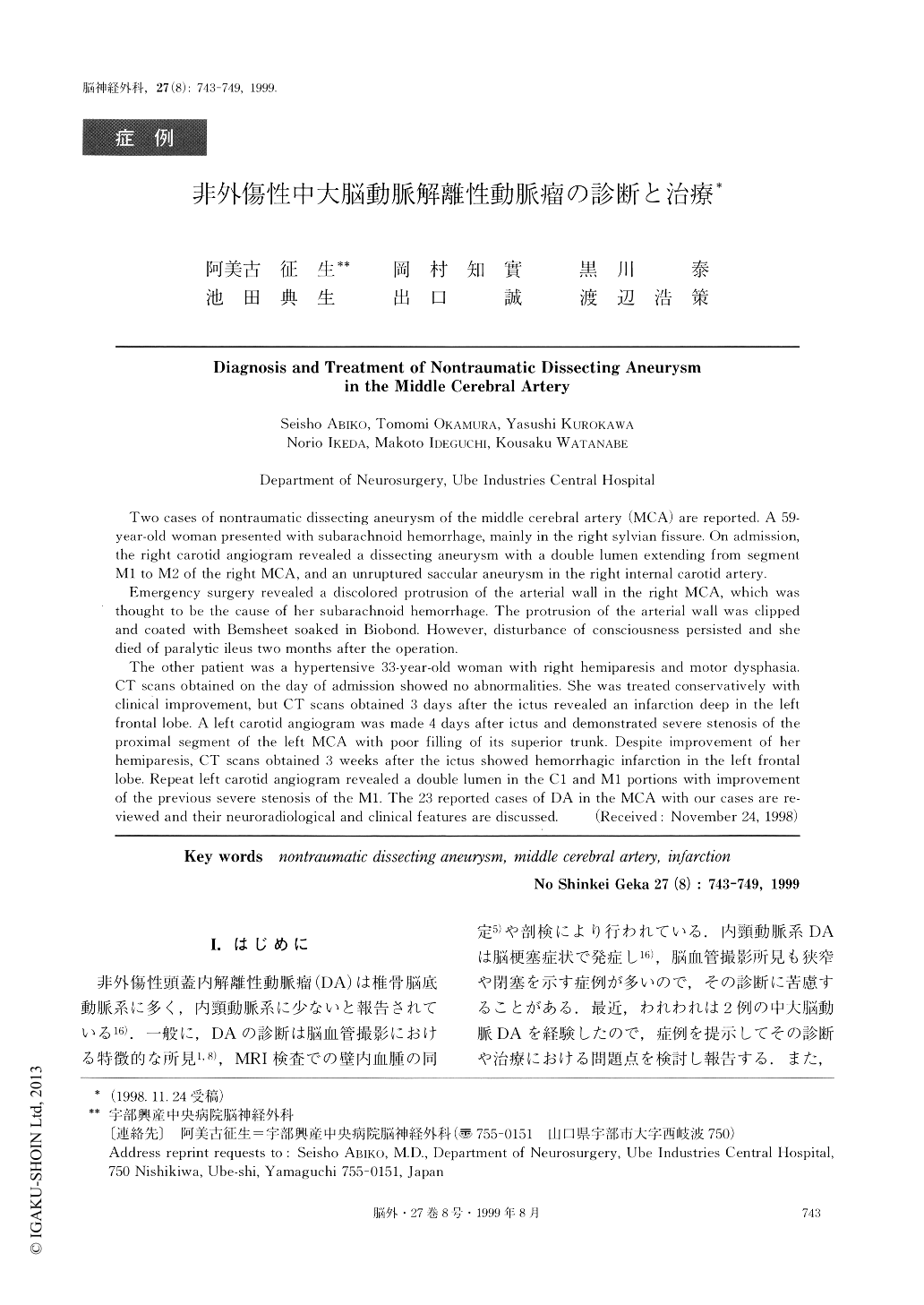
Two cases of nontraumatic dissecting aneurysm of the middle cerebral artery (MCA) are reported. A 59-year-old woman presented with subarachnoid hemorrhage, mainly in the right sylvian fissure. On admission,the right carotid angiogram revealed a dissecting aneurysm with a double lumen extending from segment M1 to M2 of the right MCA, and an unruptured saccular aneurysm in the right internal carotid artery. Emergency surgery revealed a discolored protrusion of the arterial wall in the right MCA, which wasthought to be the cause of her subarachnoid hemorrhage. The protrusion of the arterial wall was clippedand coated with Bemsheet soaked in Bioboncl. However, disturbance of consciousness persisted and shedied of paralytic ileus two months after the operation.
The other patient was a hypertensive 33-year-old woman with right hemiparesis and motor dysphasia.CT scans obtained on the clay of admission showed no abnormalities. She was treated conservatively withclinical improvement, but CT scans obtained 3 days after the ictus revealed an infarction deep in the leftfrontal lobe. A left carotid angiogram was made 4 days after ictus and demonstrated severe stenosis of theproximal segment of the left MCA with poor filling of its superior trunk. Despite improvement of herhemiparesis, CT scans obtained 3 weeks after the ictus showed hemorrhagic infarction in the left frontallobe. Repeat left carotid angiogram revealed a double lumen in the C1 and M1 portions with improvementof the previous severe stenosis of the MI. The 23 reported cases of DA in the MCA with our cases are re-viewed and their neuroradiological and clinical features are discussed.

Copyright © 1999, Igaku-Shoin Ltd. All rights reserved.


