Japanese
English
- 有料閲覧
- Abstract 文献概要
- 1ページ目 Look Inside
- サイト内被引用 Cited by
胃が嵌入部あるいは夾鞘部として関与する胃重積症はきわめて稀な疾患で,1888年Chiariにより初めてその剖検例が報告された.1946年Hobbs & Cohenは,45例を文献的に集めて報告し,考察を加えているが,本邦ではわれわれが調べえた範囲では水野,磯貝,山中,増田らの4手術治験例の報告を見るに過ぎない.最近われわれは,胃体部に対称性にみられたⅠ型早期多発胃癌による胃重積症の1例を経験し,選択的腹腔動脈造影にて興味ある所見を得たので,診断の面でいささかの考察を加えて報告する.
A 60 year old male was admitted to Sasebo Municipal Hospital on September 30, 1971, because of increasing bouts of vomiting, abdominal pain and remarkable weightloss. Family history and past history were unremarkable.
Physical examination on admission revealed that the abdomen was slightly distended with a palpable epigastric mass which was oval in shape and measuring 6 cm in diameter. The epigastrium was also slightly tender. Positive laboratory findings were anemia with RBC of 2.82 million and Hb. of 9.0 g/dl and also hypoproteinemia with serum protein of 5.1 g/dl.
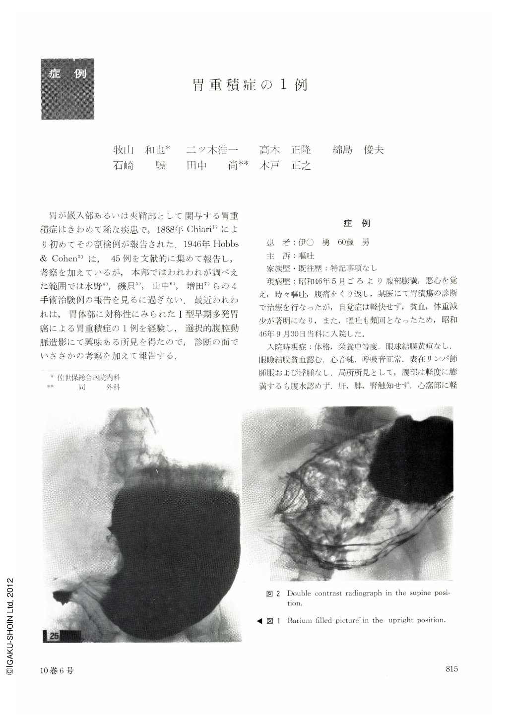
Upper GI series showed obstruction at the level of lower portion of the gastric body with resultant loss of gastric angle. The gastric antrum and the duodenal bulb could not be filled with the contrast medium. By double contrast method, several longitudinal folds of the gastric mucosa were visualized at the stenotic region.
Selective abdominal angiography was performed with the following findings. No gas shadow was seen in the anal portion of the stomach. The gastroduodenal artery originated directly from the celiac artery. The pancreaticoduodenal artery was without compression, elongation and malignant stain. Right gastro-epiploic artery ran with a significant distance from the gas shadow in the stomach.
The patient was operated on with a provisional diagnosis of submucosal tumor of the stomach. On laparotomy, the stomach was invaginated at the level of gastric body with gastro-gastro-duodenal intussusception. Palpation disclosed two polyps in the duodenum. The polyps were pedunculated and situated in the gastric body. The one at the anterior wall and the other at the posterior wall measured 5 cm and 6 cm in diameter, respectively. Histologically, these polyps were papillo-tubulary adenocarcinoma. They were also classified as type Ⅰ early gastric cancer.
Copyright © 1975, Igaku-Shoin Ltd. All rights reserved.