Japanese
English
- 有料閲覧
- Abstract 文献概要
- 1ページ目 Look Inside
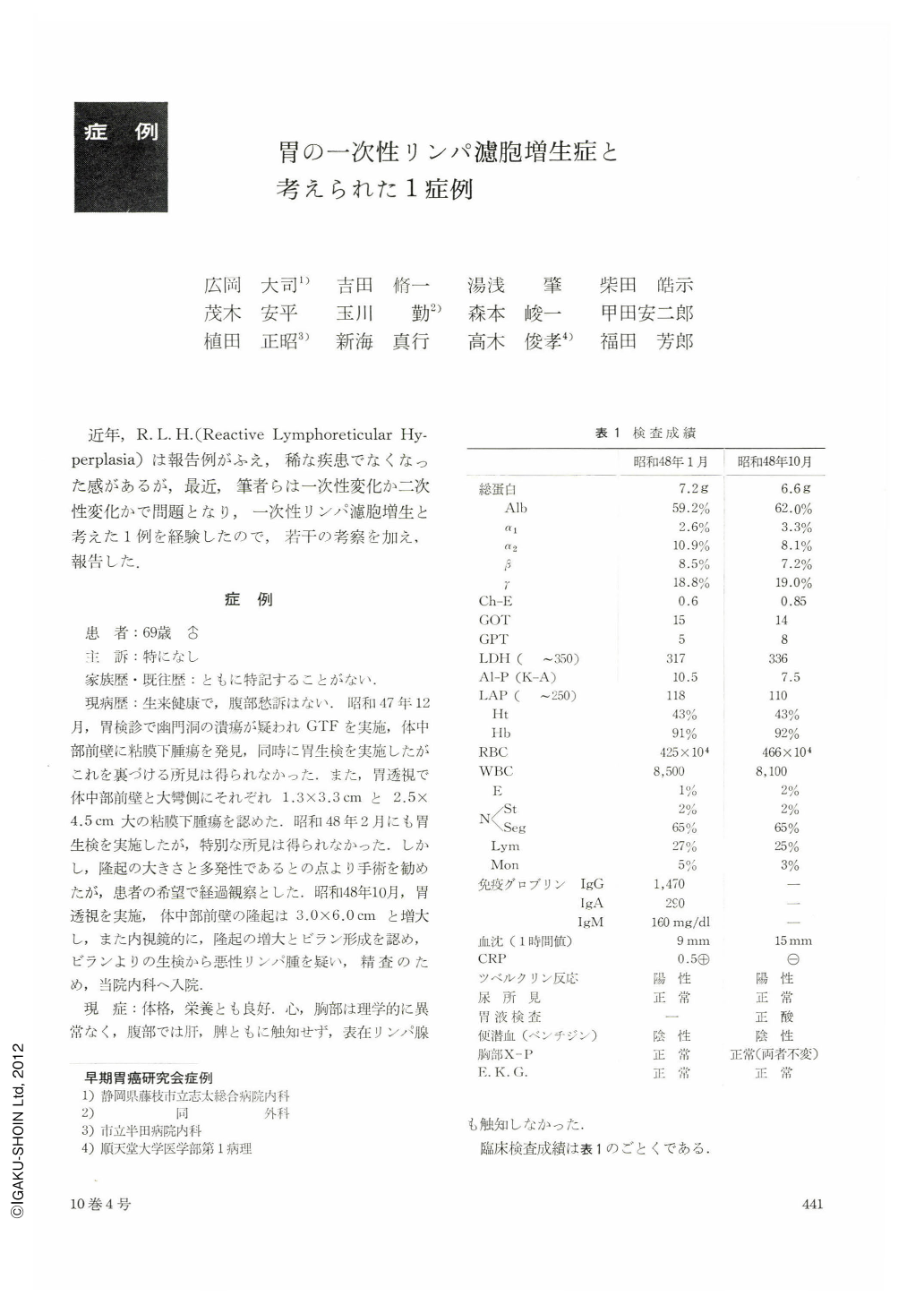
近年,R. L. H.(Reactive Lymphoreticular Hyperplasia)は報告例がふえ,稀な疾患でなくなった感があるが,最近,筆者らは一次性変化か二次性変化かで問題となり,一次性リンパ濾胞増生と考えた1例を経験したので,若干の考察を加え,報告した.
Although free from any complaints, a 69-year-old man underwent gastric x-ray check-up. As two submucosal tumors were found then at different places, the patient was placed on follow-up for 10 months long.
Roentgenological findings during this period can be summarized as follows:
1. Hypertrophy of the mucosal folds was seen on the greater curvature side from the middle to upper segments of the corpus. Also were seen findings suggestive of submucosal tumors on the anterior wall of the mid-body and likewise on the anterior wall of the upper body in the side of the greater curvature.
2. No ulcer was recognized, but“fine reticular changes associated with formation of pale erosions”were seen on the surface of the mucosal folds in the greater curvature side, that were elevated and hypertrophied.
3. The elevation on the anterior wall of the midbody grew about twice as much in size in the course of ten months. Erosions were seen on the surface as well. Altogether these findings were regarded as proliferative changes.
In view of the above-listed findings we were unable to decide whether the lesions represented reactive lymphoreticular hyperplasia (RLH) or malignant lymphoma.
Endoscopic findings were suggestive of association of a submucosal tumor with giant folds, but biopsy from the eroded surface of the elevation on the anterior wall of the mid-body led us to suspect malignant lymphoma of the stomach. Surgical intervention was accordingly performed.
The gross specimen of the resected stomach showed convolutional hypertrophy of the mucosal folds, but there was no definite evidence of ulcer. Histologically the lesions were multiple, extending over wide area with partial infiltrative proliferation. Cytologic study revealed breakdown of lymph-follicle structure, with lympho reticular tissue rather more in juvenile state in addition to giant cells and mitosis. These findings were not conclusive of the existence of malignant lymphoma, and we are more in favor of primary hyperplasia of lymph follicles than of RLH.
Copyright © 1975, Igaku-Shoin Ltd. All rights reserved.