Japanese
English
- 有料閲覧
- Abstract 文献概要
- 1ページ目 Look Inside
- サイト内被引用 Cited by
X線診断や内視鏡診断技術の著しい進歩に伴って,早期胃癌をはじめ胃内の微細病変が多数発見されるようになってきた.今回のテーマ“良・悪性の境界領境病変”も,診断技術の進歩と共にクローズアップされてきたと言ってよかろう.この病変は,異型上皮1)~4),Ⅱa-subtype5)6)など種々な名称で呼ばれているが,一般には異型上皮と呼ぶ場合が多いようである.異型上皮は,組織学的に“良性病変”でもなく,また“癌”でもない“境界領域”に属する病変であると佐野は述べている7).
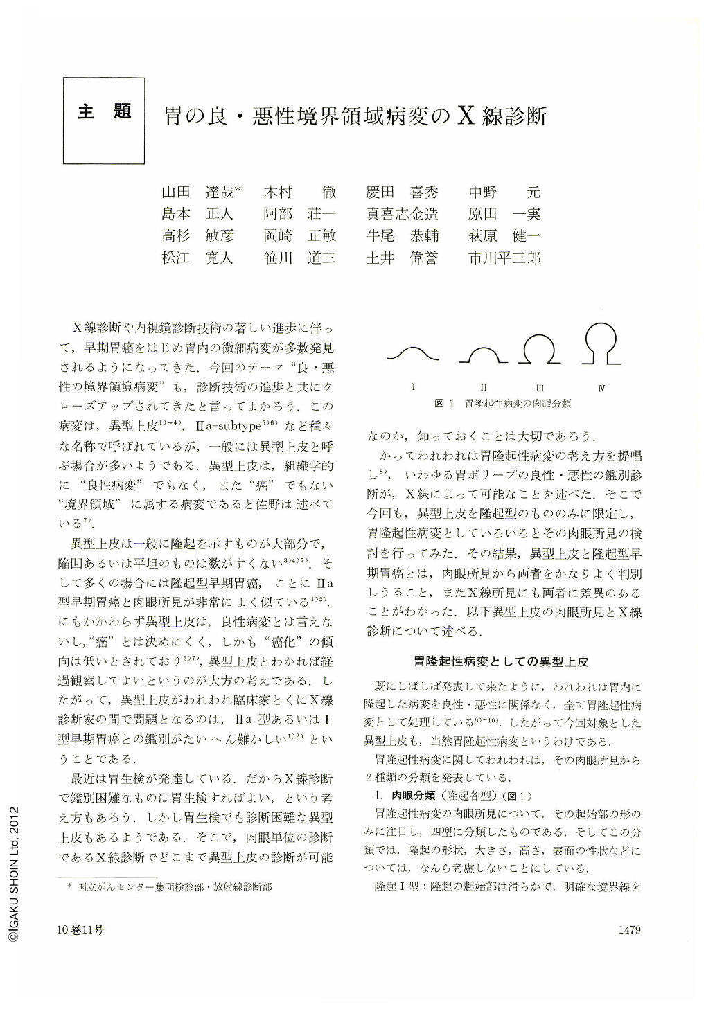
異型上皮は一般に隆起を示すものが大部分で,陥凹あるいは平坦のものは数がすくない3)4)7).そして多くの揚合には隆起型早期胃癌,ことにⅡa型早期胃癌と肉眼所見が非常によく似ている1)2).にもかかわらず異型上皮は,良性病変とは言えないし,“癌”とは決めにくく,しかも“癌化”の傾向は低いとされており3)7),異型上皮とわかれば経過観察してよいというのが大方の考えである.したがって,異型上皮がわれわれ臨床家とくにX線診断家の間で問題となるのは,Ⅱa型あるいはⅠ型早期胃癌との鑑別がたいへん難かしい1)2)ということである.
Atypical epithelium of the stomach, a borderline lesion benigancy and malignancy, has come to attract a great deal of attention along with the progress made in diagnostic techniques for gastric diseases. Mostly it shows elevated type of early gastric cancer as seen macroscopically. It is no wonder that what is most important in x-ray diagnosis of atypical epithelium is its differentiation from elevated type of gastric cancer.
Atypical epithelium of the stomach that shows elevation is naturally included in what we have advocated as elevated lesions of the stomach. As the bases of X-ray diagnosis, macroscopical findings of all elevated lesions of the stomach have been studied, including atypical epithelium and other elevated lesions of the stomach, both benign and malignant. As a result we have found out that in type Ⅱ and Ⅲ elevated cancers atypical epithelium is recognized in those less 19 mm in diameter and whose surface features are slighter than C5, or “showing relatively regular surface features”. In elevated type of early carly cancer the majority of atypical epithelium are seen in those over 20 mm in diameter and whose surface features are over C6, or “showing relatively irregular unevenness”. In the past differentiation between atypical epithelium and elevated type of early cancer of the stomach has been considered difficult both macroscopically and in roentgenogram. However, our present study seems to indicate that X-ray diagnosis of atypical epithelium is within easy reach if each type of elevated lesions of the stomach (types Ⅰ~Ⅳ) is well delineated along with its size and surface characteristics.
Some illustrative cases of atypical epithelium of the stomach are also presented together with its macroscopic features and X-ray diagnosis.
Copyright © 1975, Igaku-Shoin Ltd. All rights reserved.