Japanese
English
- 有料閲覧
- Abstract 文献概要
- 1ページ目 Look Inside
- サイト内被引用 Cited by
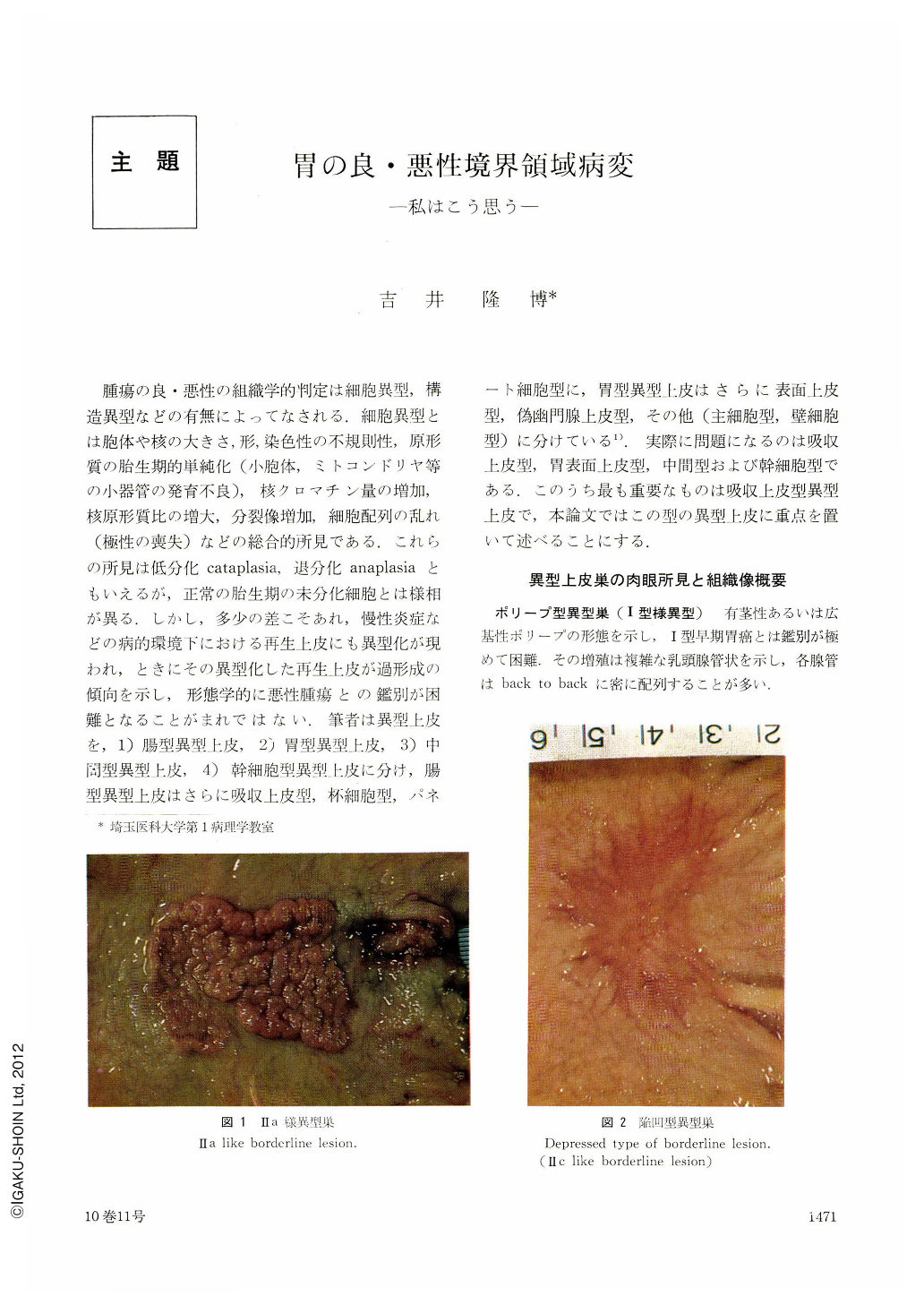
腫瘍の良・悪性の組織学的判定は細胞異型,構造異型などの有無によってなされる.細胞異型とは胞体や核の大きさ,形,染色性の不規則性,原形質の胎生期的単純化(小胞体,ミトコンドリヤ等の小器管の発育不良),核クロマチン量の増加,核原形質比の増大,分裂像増加,細胞配列の乱れ(極性の喪失)などの総合的所見である.これらの所見は低分化cataplasia,退分化anaplasiaともいえるが,正常の胎生期の未分化細胞とは様相が異る.しかし,多少の差こそあれ,慢性炎症などの病的環境下における再生上皮にも異型化が現われ,ときにその異型化した再生上皮が過形成の傾向を示し,形態学的に悪性腫瘍との鑑別が困難となることがまれではない.筆者は異型上皮を,1)腸型異型上皮,2)胃型異型上皮,3)中間型異型上皮,4)幹細胞型異型上皮に分け,腸型異型上皮はさらに吸収上皮型,杯細胞型,パネート細胞型に,胃型異型上皮はさらに表面上皮型,偽幽門腺上皮型,その他(主細胞型,壁細胞型)に分けている1).実際に問題になるのは吸収上皮型,胃表面上皮型,中間型および幹細胞型である.このうち最も重要なものは吸収上皮型異型上皮で,本論文ではこの型の異型上皮に重点を置いて述べることにする.
Borderline atypical epithelial ceh_s can be classified into intestinal, gastric, intermediate and stem cell type. Intestinal type is further classified into absorptive cell, goblet cell, paneth cell type; gastric type into surface cell, pyloric cell type and others (such as chief cell type). This paper mainly deals with absorptive cell type.
Borderline lesions are characterized by the following morphology: in cytoplasm, 1) highly columnar cell body, 2) no conspicuous irregularity in size and arrangement, 3) no mucinous granules, as a rule, 4) development of striated border in various grades, 5) basophilic tendency in stainability; in nucleus, 1) long oval shape, 2) large nucleo-cytoplasmic ratio, 3) sitting at the bottom of cytoplasm, 4) preservation of polarity, 5) uniformity in size and shape, 6) hyperchromatism, 7) finely granular chromatin distributed diffusely in nucleus, 8) small nucleolus. Conditions to differentiate carcinoma from borderline atypism are: 1) presence of irregularity in size and shape of cell body and nucleus, 2) increase of nuclear size (increase of nucleo-cytoplasmic ratio), 3) increase in amount of chromatin, 4) coarsening of chromatin granules with intensified stainability, 5) thickening of nuclear membrane (light-microscopically), 6) enlargement of nucleolus, 7) stratificatification in nuclear arrangement (migration of nuclei from the bottom to the apical portion of cytoplasm), 8) declining of polarity, 9) increase of structural atypism.
In diagnosis of malignancy, one or more conditions described above should be added to morphology of borderline atypism. However, even benign atypical cells frequently exhibit one or more conditions for malignancy. On the other hand, there are cases of carcinoma hardly demonstrating conditions for malignancy. In other words, atypism is overlapped in benign and malignant cells.
From studies with electron-microscopy, histochemistry, microspectrophotometric measurement of nuclear DNA,3H-thymidine autoradiography as well as lightmicroscopy, it can be considered that borderline atypism is in the intermediate between the benign and malignant, morphologically and biologically.
Copyright © 1975, Igaku-Shoin Ltd. All rights reserved.