Japanese
English
- 有料閲覧
- Abstract 文献概要
- 1ページ目 Look Inside
欧米の報告では,脂肪腫は消化管の中で大腸に好発し,大腸良性腫瘍の中では腺腫性ポリープに次いで多いといわれている.本邦では消化管,ことに大腸の脂肪腫はきわめて稀で,その報告例は自験例も含め23例にすぎない.最近われわれは本症例を経験したので,本邦報告例とあわせ,症状,診断,治療につき考察したいと思う.
A, forty nine-year-old female visited our hospital because of upper abdominal pain of four months' duration. The pain was related to the ingestion of food and was relieved spontaneously by not eating. It gradually increased in severity and the cramping pain in the right lower abdomen, which radiated to the right lower back, appeared. But she had no nausea and vomiting. Twice in the previous two days she had noted bright red blood at defecation. She had lost about 8 kg in weight in the past four months. It was attributable to the lack of eating for fear of bringing on an attack.
Physical examination revealed that the abdomen was soft and mildly distended with marked tenderness in the right lower quadrant. Perstaltic sounds were normal. No masses could be palpated in the abdomen. Rectal digital examination was normal.
Laboratory studies were normal except the positive occult blood of feces.
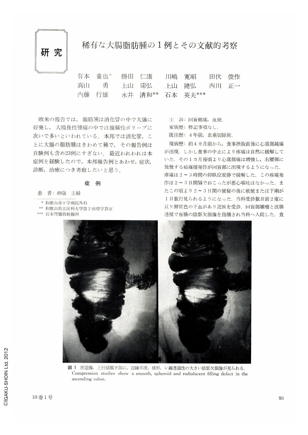
Roentogenologic examination of the colon revealed on compression studies a smooth, spheroid radiolucent filling defect measuring about 5 cm in diameter in the ascending colon just distal to the caecum. Air contrast studies showed that the tumor was pedunculated and ovoid which was producing intussusception. The impression was that this tumor was a benign one, most likely a lipoma.
The abdomen was opened and a soft freely movable mass about the size of an egg was palpated in the proximal ascending colon. There was no evidence of intussusception. There were no lymph node swelling in the mesentery. An end-to-end anastomosis was performed between the ascending colon and terminal ileum.
The gross description of the lesion was that of a pedunculated submucosal tumor measuring 4 cm in diameter. There were some loss of mucous membrane at the top and base of the tumor. On transverse section of the mass it was seen to be composed of bright yellow fatty tissue.
Microscopic description showed that the mass was composed of adult fatty cells and was situated directly beneath the muscularis mucosae. The overlying mucosa was absent on the top and base of the mass. There was prominent infiltration of the plasma cells and lymphocytes at the base with no evidence of malignancy.
The patient had an uneventful recovery with complete relief of symptoms.
Copyright © 1975, Igaku-Shoin Ltd. All rights reserved.