Japanese
English
- 有料閲覧
- Abstract 文献概要
- 1ページ目 Look Inside
カルチノイドはセロトニン分泌腫瘍として知られ,近年その報告例は増加しているが,消化管カルチノイドのうち胃原発カルチノイドの報告例は少ない.われわれは胃集団検診にて胃底部の巨大な腫瘤を発見し,手術後胃カルチノイドと判明した1例を経験したので報告し,若干の文献的考察を加えたい.
症例
患 者:佐○木○郎 49歳 男子(大工)
家族歴:父親が胃癌て死亡(69歳).母親は健在.心筋硬塞の既往がある.
既往歴:21歳の時黄疸,40歳て脳塞栓といわれ入院加療.
現病歴:昭和47年8月に心窩部不快感が出現し,9月には空腹時の心窩部痛があり,近医にて投薬をうけ軽快している,10月頃より易疲労感と食欲減退があるのて,10月27日職場て実施された胃集団検診を受け異常を指摘された.11月1日胃精密検査にて胃底部腫瘤を発見され11月10日入院した.入院時三くに自覚症状はなく,顔面潮紅,flushing,喘息様発作などのカルチノイド症候群を思わせる症状はなにもなかった.
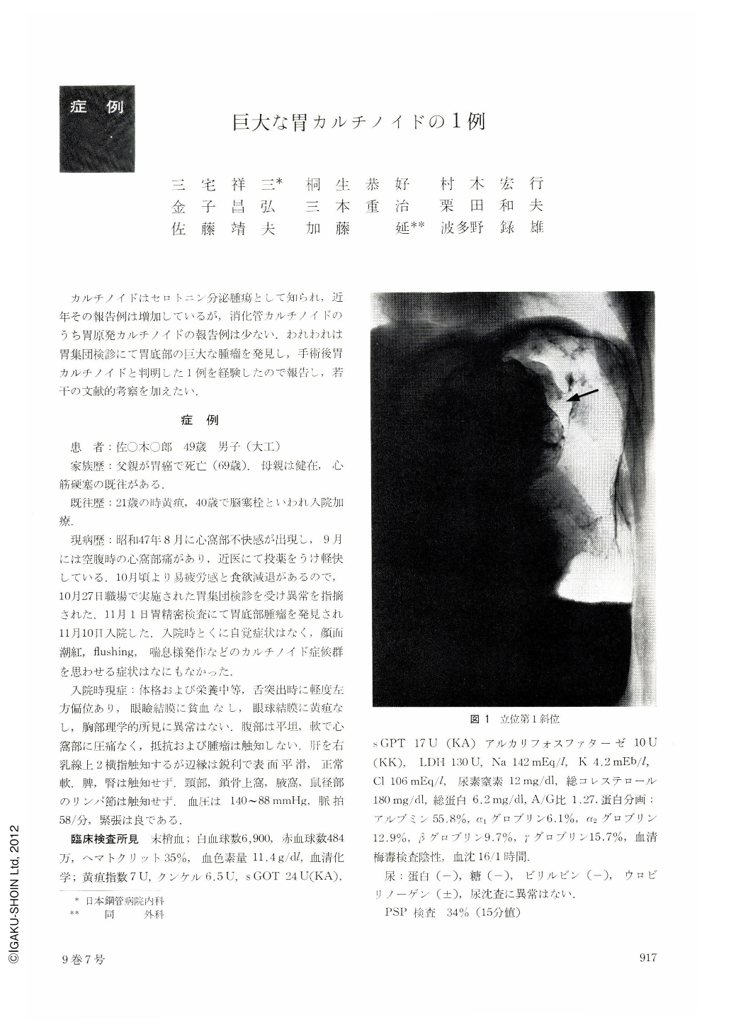
A 49-year-old male patient was found to have a large gastric tumor at a mass screening of the upper G. I. tract. The patient was admitted to our hospital, as he had complaints of abdominal distress and slight anorexia. Laboratory and physical examinations showed no abnormalities. X-ray film of the stomach revealed a giant polypous tumor protruding from the fornix of the posterior gastric wall. The tumor was confirmed as a greyish brown, giant polypous one by the gastroscopic examination performed shortly after the x-ray examination. Total gastrectomy was carried out under a diagnosis of stomach carcinoma.
The neoplasm was found in the resected stomach as a fungating spherical tumor with coarse and irregularly nodular surface, measuring 9×9×6 cm in size. Histopathological examination revealed that the tumor showed invasion mainly to the submucosal layer with bartial extension to the subserosal layer. The tumor cells, with nuclei relatively small and uniform in size, were scarce in cytoplasm, showing ribbon-like arrangement and pseudo-rosette formation. However, a small portion of the tumor cells, especially that located near the surface of the mass, showed relatively large, irregular oval nuclei having a few large nucleoli. These cells showed moderate atypism as well as several mitotic figures. Argyrophil reaction was positive by Siver-Munger stain, but Argentaffin reaction was negative by Masson-Fontana stain. The staining for mucin by PAS and Mucicarmin regents was negative. Electron microscopic examination was done using the formalin fixed material, which showed relatively small-sized, electron dense granules in the cytoplasm of the tumor cells. Thus, the tumor was confirmed as carcinoid tumor by the above pathomorphological findings.
The measurement of 5-HT in the blood and 5-HIAA in the urine was done twice, but only postoperatively. The result of the former was 5.7 mcg/dl and 4.8 mcg/dl (Udenfreind's method. normal value: 10~30 mcg/dl) and that of the latter was 4.7 mg/ 24 hrs and 3.9 mg/24 hrs (Udenfreind's method. normal value : 2~9 mg/24 hrs).
Our statistical review of cases up to the date (1972) revealed that this case was the 20th carcinoid tumor originating from the stomach reported in Japan so far.
Copyright © 1974, Igaku-Shoin Ltd. All rights reserved.