Japanese
English
- 有料閲覧
- Abstract 文献概要
- 1ページ目 Look Inside
はじめに
消化管のカルチノイドの報告は,最近比較的多くみられるようになったが,胃原発カルチノイドの症例は比較的少ない.欧米諸国では,Askanazy(1923)1)が剖検で偶然に発見して以来,1968年までに89例を数えるといわれる2).本邦における胃カルチノイドは,1962年の大杉らの報告3)を初めとして1970年までに14例が報告されており,その他,阿部によると,5例みられるという24).しかし本症例のように,術前に診断し得たのは,八尾らの報告16)に次ぐものであり,また本邦の報告症例の中では最年少であり,かつ最も小さいものであるので,経過の概要を述べるとともに,若干の文献的考察を加えてみたいと思う.
Carcinoid of the stomach is a very rare disease. In Europe and in the United States there have been only about 100 cases since Askanazy's report in 1923, while in this country only 14 such cases have been found since Osugi's initial report. The case presented here is therefore believed to be the 15th in Japan, being the second preoperatively diagnosed case of gastric carcinoid next to Yao's report. It is also of the smallest and seen in a relatively younger person.
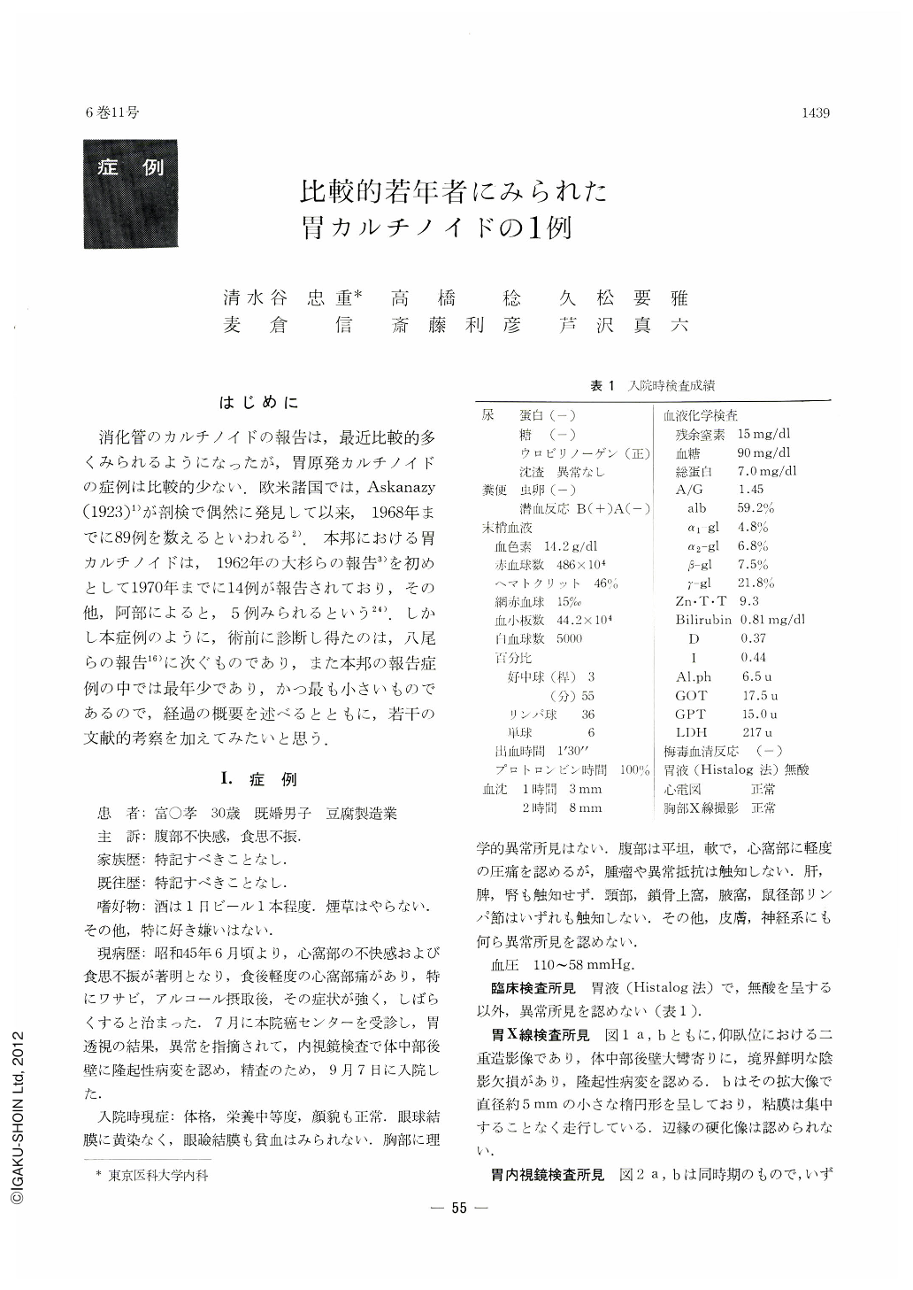
The patient is a 30-year-olcl man having chief complaints of abdominal distress and loss appetite. Except for anacidity in the gastric juice, there was nothing abnormal in the laboratory examinations.
X-ray study of the stomach revealed a small shadow defect near the greater curvature on the posterior wall of the corpus, while in gastrocamera photographs a small protruding lesion was seen at the same site, with ill-defined constriction around it and its surface not quite different from the surrounding mucous membrane. Its tip was engorged as if in an erosive state. Biopsied specimen disclosed histologically a circumscribed tumor tissue extending from the deeper part of the mucosa way through to the muscle layer. The tumor cells, with their nuclei relatively small and uniform in size, were scarce in cytoplasm, showing ribbon-like arrangement and lace-like pattern.
The quantative measurement of 5-H.T. in the blood and 5-H.I.A.A. in the urine was 0.171μg(γ)/ml average and 9.118 mg/day, respectively, showing normal account. An area about 3cm in diameter with the tumor in its center was removed. The neoplasm, measuring 5×5×2 mm, was an ill-defined hemispheric protrusion of slightly irregular surface, which was at once engorged and discolored. Histopathological findings almost coincided with those of biopsied specimen and the tumor was confirmed as carcinoid. Both mucus stain and Fontana's stain were negative.
Gastric carcinoid in its early stage, when there is neither any characteristic sign of it, nor are there any abnormal findings in biochemical examinations such as 5-H.T. in the blood anti 5-H.I.A.A. in the urine, is very difficult to arrive at its diagnosis. The case described in this paper is another illustration of the importance of biopsy under the guidance of endoscopy.
Copyright © 1971, Igaku-Shoin Ltd. All rights reserved.