Japanese
English
- 有料閲覧
- Abstract 文献概要
- 1ページ目 Look Inside
- サイト内被引用 Cited by
1.はじめに
胃疾患に対する各種診断の驚異的進歩はここ数年間めざましく,早期胃癌として診断する病変が漸次小さいものを対象とするようになったのは自然のなりゆきであった.とくに1966年の日本内視鏡学会秋季大会では,直径2cm以下の表面型早期胃癌を微細病変と呼んで,この微細病変の診断およびその限界がとりあげられ検討された.さらに1969年の第7回日本内視鏡学会秋季大会では,1cm以下の小胃癌(微小胃癌)を対象として検討を加えるまでにいたり,微細病変として直径2cm以下のものから,ついに1cm以下の小病変の診断となって,診断の限界も肉眼的にぎりぎりの線まで到達して早期胃癌の診断進歩のすさまじさを物語っている.2cm以下の微細病変としてとりあつかった胃癌の診断にあたっても,肉眼的単位の診断学は良性,悪性の質的診断が困難であることは,すでに発表したが8),肉眼的診断として病変の存在診断に,細胞診,胃生検による組織学的,質的診断が加わって存在診断と質的診断の両者を必要としてきている.
直径1cm以下の小胃癌の診断にあたって,その病変の存在を肉眼的に検討して,その大きさの診断の限界について言及してみたい.
In recent years tremendous strides have been made in various diagnostic technics for diseases of the stomach. In fact more minute lesions have come to be the subject of study in the diagnosis of early gastric cancer. While at the Endoscopic Meeting for Gastric Cancer in 1966 superficial type of early gastric cancer under 2cm in diameter was defined as minute cancer lesion and its diagnosis together with its diagnostic limits was discussed, the subject of investigation at the same Meeting in 1969 was narrowed down to minute gastric cancer under 1 cm in diameter.
This paper deals with the macroscopic diagnosis of minute gastric cancer under lcm in diameter with special reference to gross investigations of its existence together with its diagnostic limits to macroscopic confirmation.
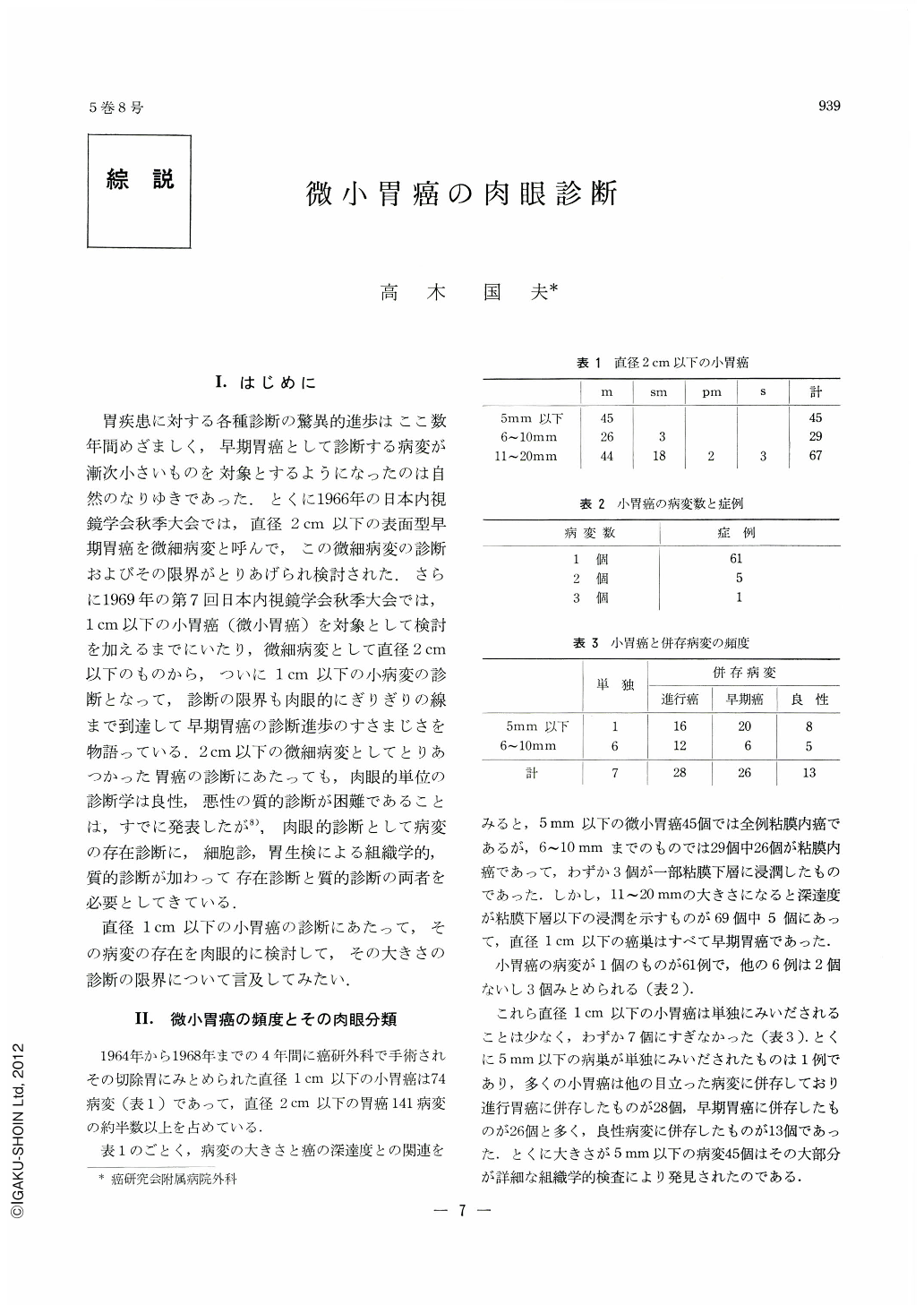
In the past four years, 74 lesions of minute gastric cancer under 1 cm in diameter were encountered in patients operated on in the Surgical Department of the Cancer Institute Hospital. Of these, 45 lesions under 5mm belonged to intramucosal cancer; partial submucosal invasion was seen only in 3 lesions out of the remaining 29 cancer lesions measuring from 6 to 10 mm in diameter. Only in 7 lesions was minute cancer solitary, and only one cancer lesion under 5mm was single. Most of minute cancer lesions were co-existent with other more striking lesions, especially with other early cancers.
Macroscopically classified, 17 lesions belonged to Ⅱa, 20 to Ⅱb and 23 to Ⅱc. All of Ⅱb were minute cancers under 5 mm in diameter.
Macroscopic grouping of early gastric cancer shows that diagnosis of its gross type is not difficult when a lesion has the diameter of more than 2cm. However, this can hardly be applied to lesions under 1cm in diameter, still less to those about 5 mm in diameter. Much remains to be done for those smaller lesions.
It also has been investigated regarding the diagnostic limits of minute cancer under 1 cm in diameter. Type I was so small in number that nothing can be said about it at present. Lesions of Ⅱa under 5mm in diameter can be recognized as polypoid ones, but no discrimination is possible between their benign and malignant natures. Lesions having the diameter of more than 6mm, however, were diagnosed either as cancer or its suspect.
Lesions of Ⅱb were all under 5mm diameter and their retrospective studies based on histological examinations were not sufficient to substantiate them in the resected stomachs. Small IIc lesions about 5mm in diameter were diagnosed either as cancer or its suspect when depression was apparent, but when it was indistinct, even lesions having the diameter of about 1cm could not be confirmed as cancer; some were diagnosed as erosions instead.
In the macroscopic diagnosis of lesions under 1cm in diameter, the minimum size of cancer lesion that can be confirmed macroscopically as such is considered to be about 5mm in diameter.
Copyright © 1970, Igaku-Shoin Ltd. All rights reserved.