Japanese
English
- 有料閲覧
- Abstract 文献概要
- 1ページ目 Look Inside
Ⅰ.はじめに
最近,胃内視鏡の発達はめざましく,とくに,胃疾患の内視鏡診断に関しては多方面から活発に研究されている.しかし,胃の病態生理に関する内視鏡的研究は不十分で,その報告も少ない.その原因のひとつとして,生きたままの胃内を観察する場合に部位的めやすがつき難いことが挙げられる.臨床上,古くから用いられている胃の各部の名称17)には,噴門,穹窿部,胃体部,胃角部,幽門部,幽門などがあるが,このうち,噴門と幽門は,胃と食道および胃と十二指腸の境い目であり,それぞれ,括約筋様の筋層構造1)17)により位置づけられている.また,内視鏡的にも狭窄部として明らかに観察できる.胃の形の上から小彎の屈曲部を胃角部と呼び,それを境いに胃体部と幽門部に区別されている.胃角部については,内視鏡的に主要な部位的めやすとして広く用いられているが,解剖組織上の裏付けはなく,体位の変換や胃運動によって可動性のものか否か,また,症例によって同一のものであるか否かを明らかにした報告は文献上みあたらない.そこで,内視鏡上の胃角部が胃筋層上のどこに位置するかについて検討した.
もうひとつ胃の組織学的な部位を表わす名称として,胃底腺幽門腺粘膜境界部8)があるが,この粘膜境界部は普通の内視鏡検査では観察し難いもので,教室の成績によれば,その存在部位は個人差が大きい.Congo-red液を用いて粘膜境界部を観察しようとする動物実験7)および臨床的研究4)6)は,かなり多く報告されてきたが,Congo-red法の評価についてはまちまちである.Congo-red液の変色の境い目の観察は,手術時に胃切開6)または漿膜面からの透見4)7)を行なう方法が多く用いられ,内視鏡的に検討した報告は少ない15).そこで,著者らは臨床例について,手術時全身麻酔下でCongo-red法を行ない,内視鏡によるCongo-red法の評価について検討した.すなわち,胃粘膜面に現われたCongo-red液の変色の境い目とその切除胃の胃底腺幽門腺粘膜境界部の位置的関係について検討した.
Among endoscopic studies of gastric diseases, those of comparison between the gastric corpus and the pyloric region are quite numerous. There exists nevertheless conflict of definition as regards the boundary between the two. Endoscopically the gastric angle is considered as its landmark, while histologically it is to be found in the fundo-pyloric mucosal boundary.
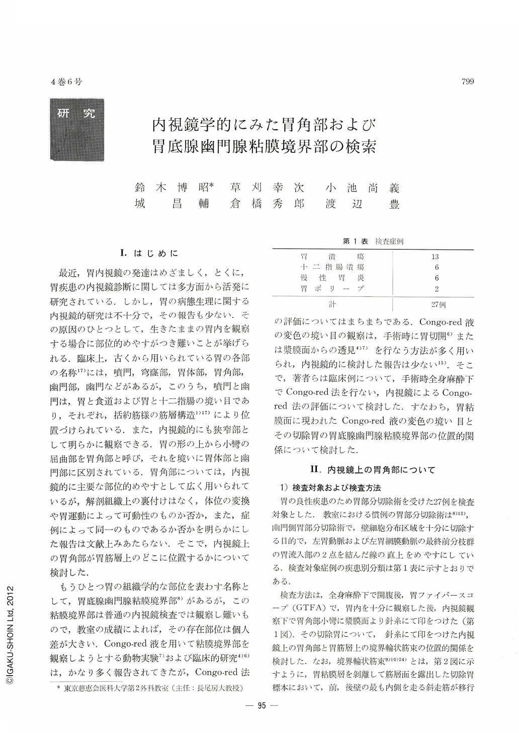
In this paper are first described the results of investigations concerning the gastric angle endoscopically considered with its relation to the architecture of adjacent muscle layers. Secondly it has been attempted whether or not it is possible to observe endoscopically the above-mentioned mucosal boundary. In all, 27 cases of partial gastrectomy were examined.
1. Gastric angle endoscopically considered.
Method of examination. During laparotomy under general anesthesia, the gastric angle endoscopically regarded as such was hooked by a threaded needle from the serosal side while observing the gastric lumen by gastroscopy. In the resected stomach, in which the gastric mucosa was stripped off so as to lay bare the muscle layers, the gastric angle (endoscopic) as was indicated by a short piece of thread remaining therein was studied as to its topographical relation with the border circular muscle bundle.
Result of examination. The gastric angle as determined by endoscopy corresponded completely with the border circular muscle bundle.
2. Fundo-pyloric mucosal boundary.
Method of examination. In the stomach of every case examined, after it had been subjected to Moe's test by Congo-red solution, a threaded needle was hooked into an edge of discoloration from the serosal side with simultaneous observation of the gastric lumen by endoscopy. That part where a piece of thread had been left behind (discolored edge) was studied as regards its topographic relation with the mucosal boundary histologically determined.
Result of examination. Of 27 cases thus examined, the discolored edge was adjacent to the mucosal boundary in 78 per cent. There was not one case in which the discolored border was located distal from the histological mucosal boundary.
Two facts thus became clear; (1) the gastric angle endoscopically considered as such corresponds with the border circular muscle bundle; (2) endoscopic observation of the fundo-pyloric mucosal boundary is feasible by Congo-red method.
Lastly some reference is made to the clinical applicatation of these facts.
Copyright © 1969, Igaku-Shoin Ltd. All rights reserved.