Japanese
English
- 有料閲覧
- Abstract 文献概要
- 1ページ目 Look Inside
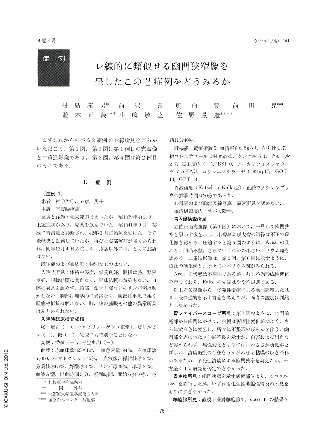
まずこれからのべる2症例のレ線所見をごらんいただこう.第1図,と二重造影像であり,第3図,第4図は第2例目のそれである.
Two cases of pyloric stenosis each of different etiology are described in this paper. Roentgenologically they looked almost alike, but postoperative histological study disclosed that one was due to atrophic lymphoblastomoid gastritis, while the other was caused by advanced cancer which developed like a Ⅱc type early cancer.
The first case is that of a 57 years old male, who began to complain of epigastric pain since two years ago. Both and x-ray endoscopic study brought out pyloric stenosis accompanied by extreme atrophic change and erosion. Biopsy was accordingly performed because multiple ulcers or Ⅱc type early cancer were suspected, but it failed to demonstrate anything but a picture of hyperplastic atrophic gastritis. At operation it was found that pyloric stenosis was caused by linear ulcer surrounding the pyloric ring. Endoscopic findings of gastritis were confirmed as those of benign atrophic lymphoblastomoid gastritis.
The second case belongs to a 43 years old female, who had persistent, though a little overstated, pain in the epigastrium. Her x-ray films closely resembled with those of the first case as far as pyloric stenosis was concerened. An extensive Ⅱc like lesion suggested by endoscopic examination proved to be mucoid cell adenocarcinoma by biopsy. At operation a superficially spreading cancer was found near the pyloric ring accompanied with cancerous infiltration into the muscularis mucosae. Pyloric stenosis was caused by it.
As for the differentiation between atrophic lymphoblastomoid gastritis and Ⅱc type early gastric cancer, one should notice that (1) in the former, multiple ulcers are apt to occur; and (2) the margins of depressed mucosa are less distinct than those of Ⅱc type lesion; (3) the surface of de pressed mucosa in the former has almost normal sheen; and (4) broken-off tips of mucosal folds look less worm-eaten as compared with those of Ⅱc type early cancer.
One should always endeavor to achieve exact diagnosis by every possible means, especially by free use of biopsy.

Copyright © 1969, Igaku-Shoin Ltd. All rights reserved.


