Japanese
English
- 有料閲覧
- Abstract 文献概要
- 1ページ目 Look Inside
- サイト内被引用 Cited by
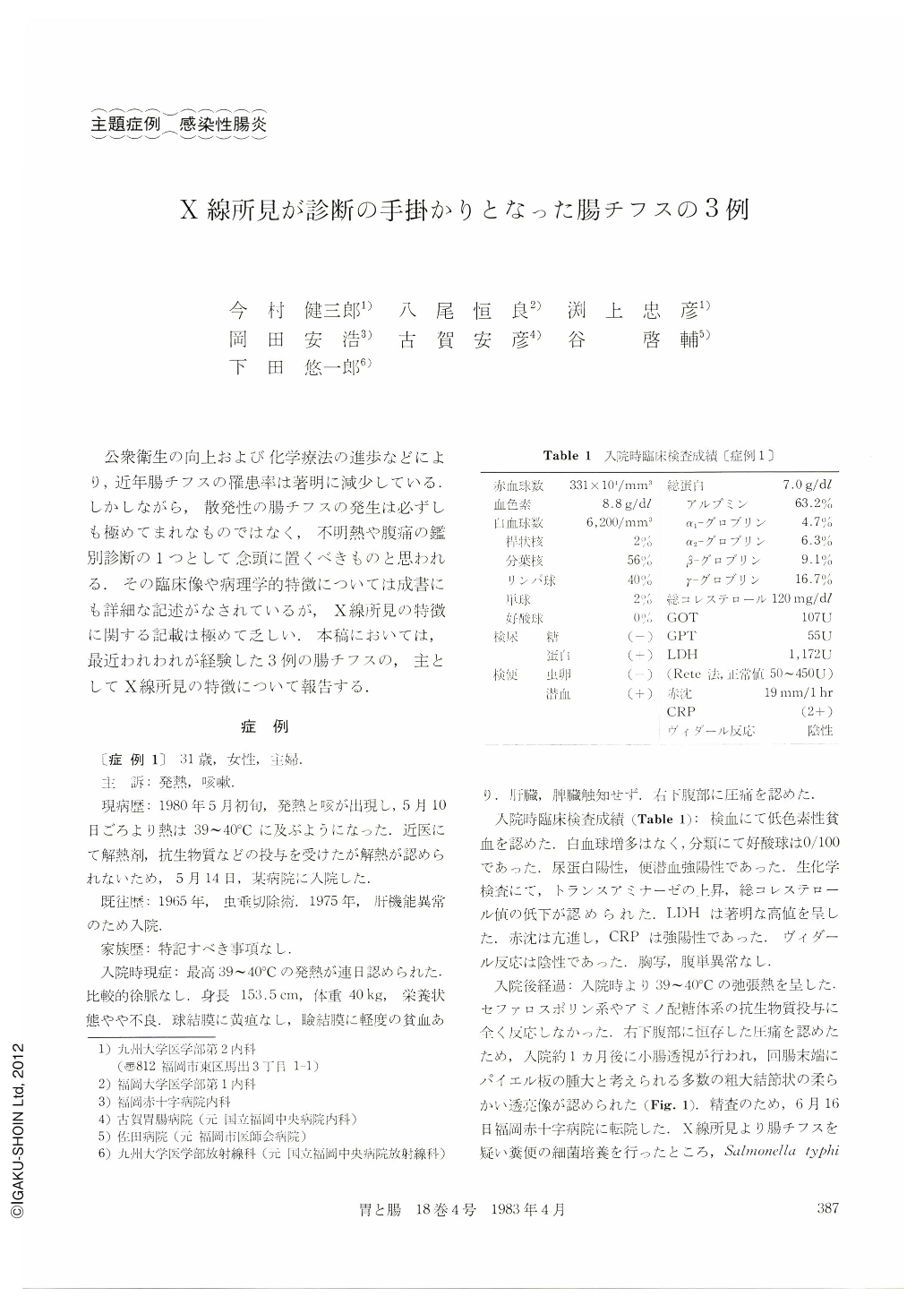
公衆衛生の向上および化学療法の進歩などにより,近年腸チフスの罹患率は著明に減少している.しかしながら,散発性の腸チフスの発生は必ずしも極めてまれなものではなく,不明熱や腹痛の鑑別診断の1つとして念頭に置くべきものと思われる.その臨床像や病理学的特徴については成書にも詳細な記述がなされているが,X線所見の特徴に関する記載は極めて乏しい.本稿においては,最近われわれが経験した3例の腸チフスの,主としてX線所見の特徴について報告する.
Two cases with Salmonella typhi infection and one with S. paratyphi B infection are reported,〔Case 1〕 was a 31 year-old woman. She was admitted to a hospital on May 14, 1980, due to high fever (39°to 40℃) which had developed four days prior to admission. In spite of accelerated ESR and strongly positive CRP, leukocytosis was not recognized. Analysis of leukocytes revealed no eosinophils, serum level of LDH was elevated, and Widal's test was negative. On x-ray examination of the small intestine, coarsely nodular pattern of the terminal ileum, presumably due to enlarged Peyer's patches was noticed. Typhoid fever was suspected and a culture of the feces was prepared. The culture was positive for S. typhi. 〔Case 2〕 was a 36 year-old man. He caught a high fever (39°to 40℃) and diarrhea on February 18, 1975. Although the fever became lowgrade after two weeks, melena newly developed on March 3. He was admitted to the National Fukuoka Central Hospital on March 11. ESR was accelerated. Leukocytosis was not recognized and there was no eosinophil on analysis. The level of LDH was within a normal range. On x-ray examination of the small intestine, coarsely nodular and serpiginous mucosal pattern of the terminal ileum was seen Suspecting infectious enteritis, a culture of the feces was prepared. It was positive for S.typhi and Widal's test was also positive. 〔Case 3〕 was a 25 year-old woman. Fever (38.2℃) developed on April 20, 1975, and continued for a week. He also caught diarrhea on April 25. She was admitted to the Fukuoka City Medical Association Hospital on April 28. ESR was accelerated. Leukocytosis was not recognized and analysis of leukocytes revealed one eosinophil out of 100 white blood cells. The level of LDH was elevated. X-ray examination of the small intestine revealed a coarsely nodular and serpiginous mucosal pattern of the terminal ileum. The culture of the feces was positive for S .paratyphi B.
All of the three cases showed similar roentgenographic findings, i.e., coarsely nodular and serpiginous mucosal patterns. It is supposed to represent swollen and enlarged Peyer's patches. This roentgenographic abnormality seems to be discriminated from that of Crohn's disease, intestinal tuberculosis, simple ulcers and lymphoid hyperplasia. Therefore, typhoid fever should be suspected when such an abnormality is noticed.
Copyright © 1983, Igaku-Shoin Ltd. All rights reserved.