Japanese
English
- 有料閲覧
- Abstract 文献概要
- 1ページ目 Look Inside
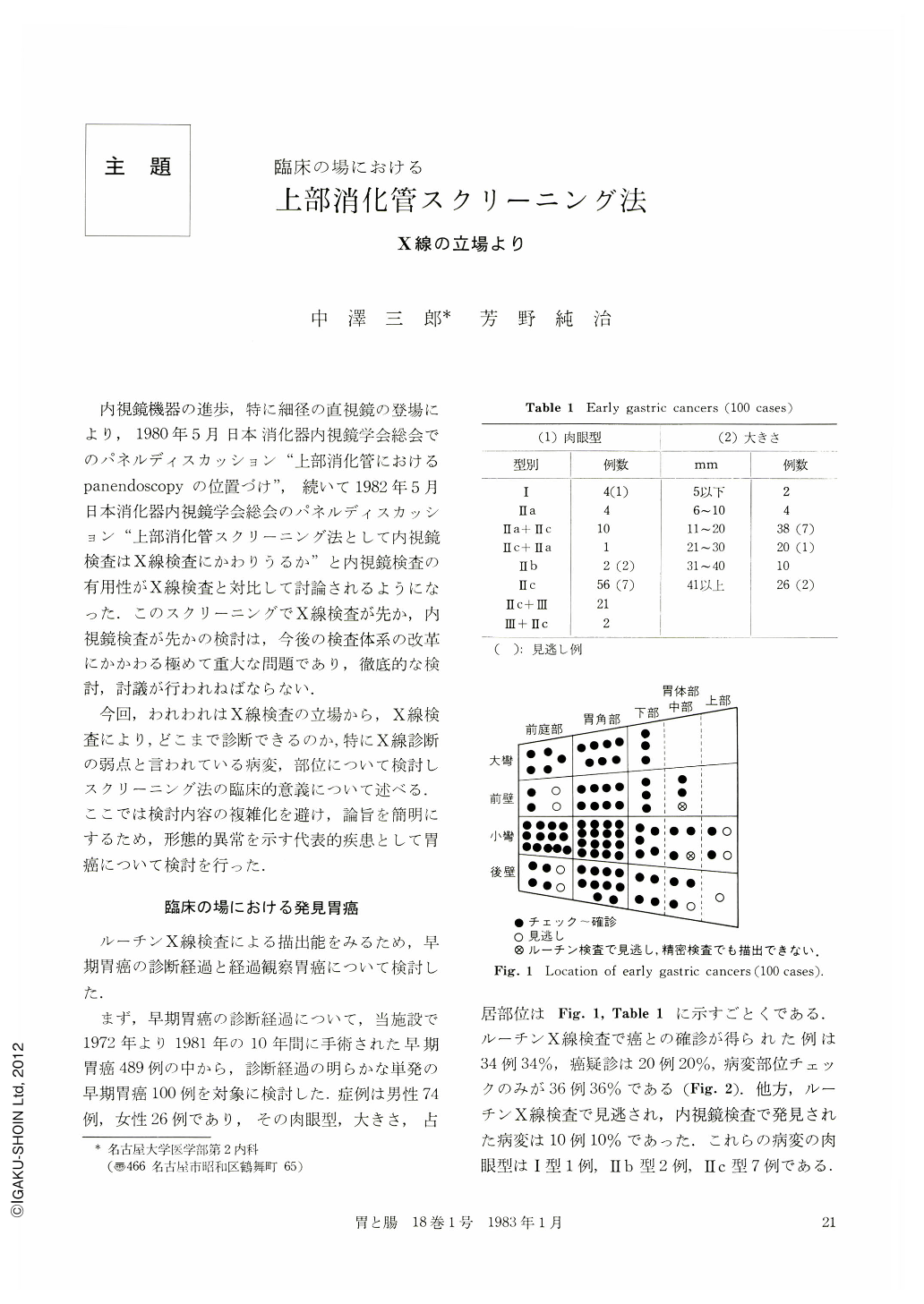
内視鏡機器の進歩,特に細径の直視鏡の登場により,1980年5月日本消化器内視鏡学会総会でのパネルディスカッション“上部消化管におけるpanendoscopyの位置づけ”,続いて1982年5月日本消化器内視鏡学会総会のパネルディスカッション“上部消化管スクリーニング法として内視鏡検査はX線検査にかわりうるか”と内視鏡検査の有用性がX線検査と対比して討論されるようになった.このスクリーニングでX線検査が先か,内視鏡検査が先かの検討は,今後の検査体系の改革にかかわる極めて重大な問題であり,徹底的な検討,討議が行われねばならない.
今回,われわれはX線検査の立場から,X線検査により,どこまで診断できるのか,特にX線診断の弱点と言われている病変,部位について検討しスクリーニング法の臨床的意義について述べる.ここでは検討内容の複雑化を避け,論旨を簡明にするため,形態的異常を示す代表的疾患として胃癌について検討を行った.
The diagnostic ability of the upper gastrointestinal lesion by means of x-ray examination was investigated in 100 cases of single gastric cancer resected from the patients in Nagoya University Hospital and the method of the routine x-ray examination executed in our clinic as stated below is reported.
By the routine x-ray examination of the upper gastrointestinal tract, 34 cases were correctly diagnosed as malignant, 20 cases were suspected as malignant, 36 cases were just suspected abnormal and 10 cases were overlooked. Those overlooked cases were one case of Ⅰ type in the cardiac area, two cases of Ⅱb type and seven cases of Ⅱc type stomach cancer.
On the other hand, 95 cases of 100 cases were correctly diagnosed by means of the more detailed x-ray examination. One lesion of the Ⅱb type and another lesion in the anterior wall in a fleshy patient were difficult to detect even by the close x-ray examination.
The follow-up cases we that were unable to detect in the initial x-ray examination were retrospectively studied to make clear the reasons of diagnostic errors. There were four lesions hidden by the contrast medium in the small intestine, six lesions existing in the folds of the gastric body, one lesion without converging folds, one lesion of Ⅱc type with shallow depression and two small cancers in the early stage of cancer, and three overlooked cases of advanced cancer in the upper gastric body.
As for the size of the gastric cancer, all lesions more than 4 mm in its maximum diameter were detectable in the gastric antrum and the gastric angle. In addition it has been reported that the lesions whose size was 2~3 mm in their diameter could be correctly diagnosed.
The improvement of the endoscopic apparatus for the upper gastrointestinal tract has brought a remarkable effect to ease the patients' pain and reduce their distress. Now, some gastroenterologists have strong confidence in the endoscopy as it is superior to the radiological examination though both relying on the screening method of the upper gastrointestinal tract. However, the endoscopy has its inevitable demerits too in a sense that there are some areas of the gastroduodenal mucosa hard to observe and its pictures are not so clear.
Accordingly it should be emphasized that the x-ray examination of the upper gastrointestinal tract still has the important roles in screening under the present circumstances.
The routine x-ray examination must be fully esteemed as long as it be made as carefully as in the detailed x-ray examination.
The method of the routine x-ray examination of the upper gastrointestinal tract in our clinic is as follows: (1) Relief picture in prone position. (2) Compression picture in upright position. (3) Barium-filled picture in upright position. (4) Barium-filled picture in prone position. (5) Double contrast picture in right oblique supine position. (6) Double contrast picture in front supine position. (7) Double contrast pictures in right lateral position, left oblique supine position, right oblique supine position, and right oblique prone position. (8) Double contrast picture in left oblique position of semi-up right posture. (9) Examination of the esophagus.
Copyright © 1983, Igaku-Shoin Ltd. All rights reserved.