Japanese
English
- 有料閲覧
- Abstract 文献概要
- 1ページ目 Look Inside
本症例は,1970年4月より1971年1月までの約9カ月間に,潰瘍の縮小と再発を認め,その初回検査時より生検による経過観察を行なったⅢ型早期胃癌の悪性サイクル例である.
患 者:石○冨○弥 35歳 男.
主 訴:空腹時心窩部痛.
既応歴:特記すべきことなし.
家族歴:特記すべきことなし.
現病歴:初診 1965年4月16日.
約5年前より,むねやけが強かったが,3週間前より食事摂取により緩解する空腹時の心窩部痛があり当院を訪れた.
A malignant cycle was seen in a case of type Ⅲ early gastric cancer followed up from its initial examination up to biopsy. In the course of 9 months from April 1970 to January 1971, ulcer first diminished in size and then it became active again.
The patient: F. I., 35-year-old male.
Past history: noncontributory.
Family history: ditto.
Present illness: initial examination on April 16, 1965.
Since about 5 years before he had complained of severs heartburn, and since 3 weeks before he had felt hunger pain with food relief in the epigastrium.
Status presens: The patient, of moderate stature, is well nourished. Has no anemia nor icterus in the conjunctivae. Heart and lung normal. The abdomen is flat and soft, but the epigastrium is tender and resistent on palpation. Liver, spleen and kidneys not palpable, nor are lymph nodes in the neck, subclavian cavities.
Laboratory examinations: red blood cells 434×104, hemoglobin (Sahli) 80%, hematocrit 38%, white blood cells, 57,000. Total protein 7.2 g/dl, A/G ratio 1.5, sedimentation rate 3 in one hour, 10 in two hours. Urine: albumin (-), sugar (-), urobilinogen (-). Sediment unremarkable. Stools were positive for occult blood by benzidine, but negative by guajac. Function tests of the liver and kidneys within normal limits. Gastric juice showed hyperchlorhydria after histamin stimulation.
Diagnostic courses: ―
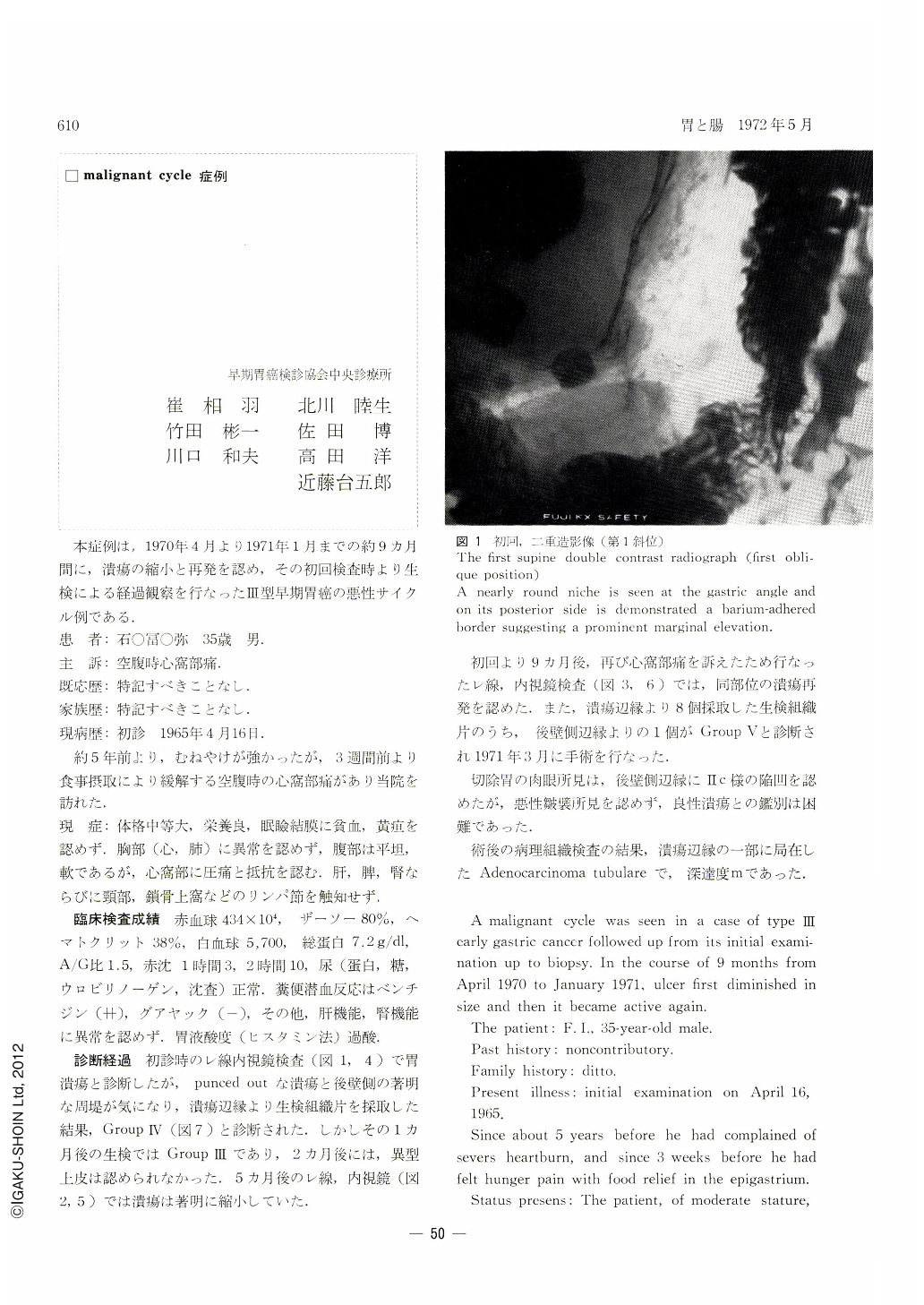
At the initial examinations by x-ray and endoscopy (Figs. 1 and 4) the lesion was diagnosed as gastric ulcer, but it was “punched out” and distinct embankment on the posterior wall side was such that tissue for biopsy was removed from the margins of the ulcer. The diagnosis then was Group Ⅳ (Fig. 7). Another biopsy one month later disclosed it as Group Ⅲ and 3 months later no atypical epithelium was seen. Pictures of x-ray and endoscopy 5 months later (Fig. 2 and 5)revealed that ulcer had been remarkably reduced in size.
Nine months after the initial examination the patient again complained of epigastric pain, so that he was re-examined by x-ray and enoscopy (Figs. 3 and 6). Ulcer had recurred at the same place. Of 8 pieces of tissue taken for biopsy from the ulcer margins, one from the border in the posterior wall side was judged Group Ⅴ. The operation was performed in March 1971.
Gross specimens of the resected stomach, although slightly depressed as in a Ⅱc lesion on the ulcer margin in the posterior wall side, did not yield any finding suggestive of malignant mucosal folds. It was quite difficult to discriminate it from benign ulcer.
Histopathological study of the excised stomach revealed adenocarcinoma tubulare localized on one side of ulcer borders, with m degree of depth invasion.
Copyright © 1972, Igaku-Shoin Ltd. All rights reserved.