Japanese
English
- 有料閲覧
- Abstract 文献概要
- 1ページ目 Look Inside
大腸早期癌の報告は胃早期癌に比べてまことに少く,術前診断例は文献上でもまれである9).St. Mark's HospitalにおいてMachida FCS & Olympus CF-LBを用いて行った112例の大腸内視鏡検査で著者らは6例の癌を発見した.そのうち2例はいわゆる早期癌であり内視鏡的に診断可能であったのでここに報告したい.
[症例1]
患 者:53歳,男.
主 訴:下血.
家族歴:特記すべきことなし.
既往歴:34歳.肺結核のため左肺切除.
現病歴:1969.10.旅行先で大量下血のため輸血,原因不明であった.
In 112 colonoscopic examinations at St. Mark's Hospital using the short and long colonoscope (Machida FCS & Olympus CF-LB) we have detected 6 examples of resectable carcinomas of the colon. This report describes two cases of early carcinoma in our series.
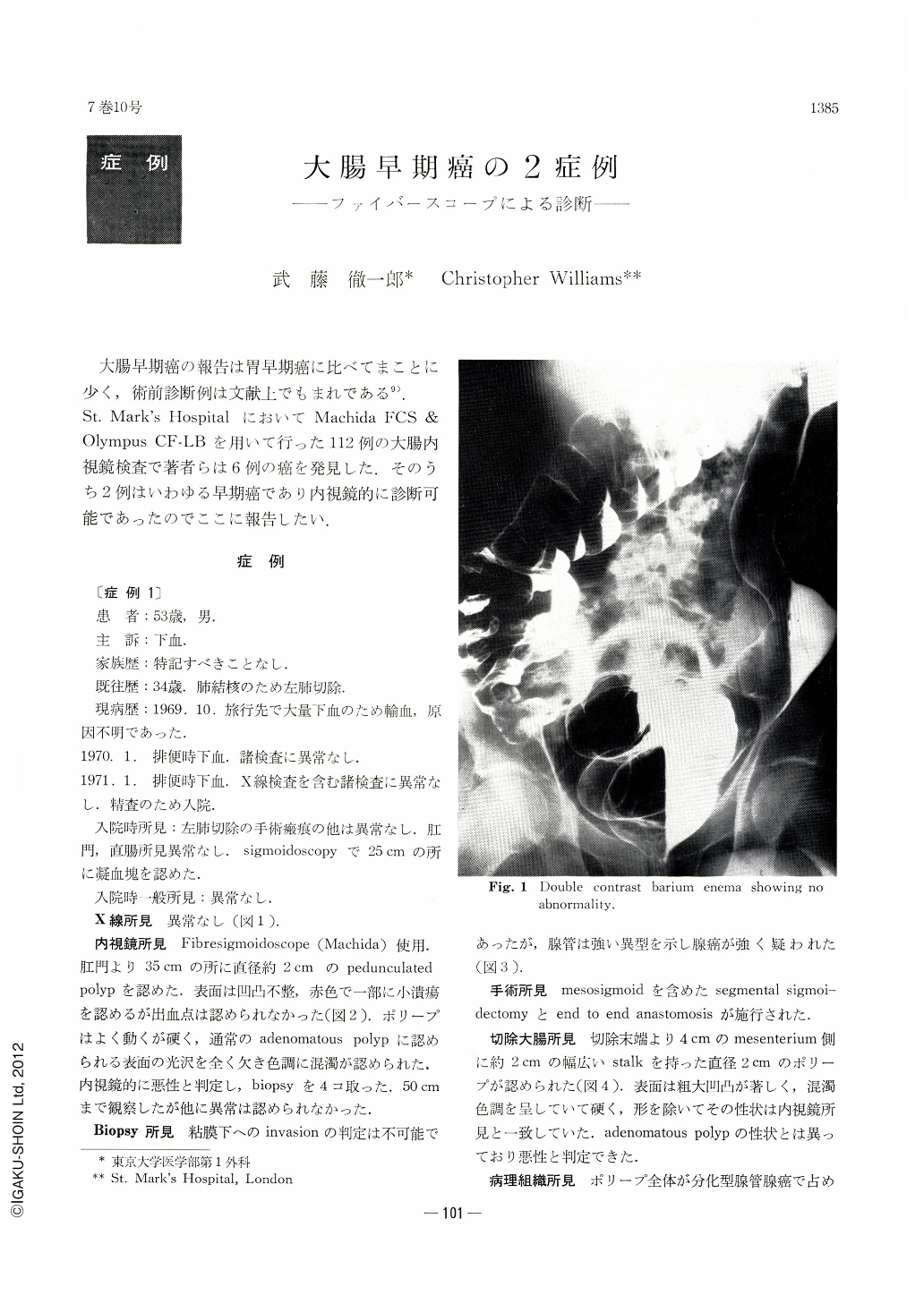
Case 1. A 53-year-old man had a one year history of rectal bleeding. The double contrast barium enema showed no abnormality. On colonoscopy a pedunculated polyp 2 cms in diameter was detected in the sigmoid colon. It had an irregular, dark-red and opaque surface with shallow ulceration and was firm in consistency, suggestive of malignancy. The biopsy showed severe epithelial dysplasia suggestive of carcinoma. The operation specimen confirmed the presence of a polyp in the sigmoid colon. Histology showed a well-differentiated adenocarcinoma of a low grade of malignancy with early invasion of the stalk.
Case 2. A 62-year-old man with multiple polyps of the colon. He had a polyp in the descending colon and 2 rectal adenomas had been removed 6 years previously. The patient was followed-up every year for 6 years until 1971 when there was an apparent increase in size of the polyp, raising the possibility of malignant change. On colonoscopy 8 polyps were detected in the colon, all but one being under 7 mms in diameter. The largest polyp in the descending colon was 1 cm in diameter on a short stalk. It had a rough, dark-pink and opaque surface with shallow ulceration and was firm in consistency, suggestive of malignancy. The biopsy showed fragments of adenoma with dysplastic epithelial proliferation but there was no evidence of invasion by carcinoma. The operation specimen confirmed the presence of a polyp in the descending colon together with 19 small polyps scattered throughout the colon. Histology showed a well-differentiated adenocarcinoma of a low grade of malignancy in an adenoma. This is a good example of an adenoma undergoing malignant. Sections of all the small polyps showed adenomas.
These two cases come into the category of early carcinoma of the colon and rectum as defined by Morson (1968). Even in operation specimens the incidence of early carcinoma is low (3.3% in 2,305 rectal cancers at St. Mark's Hospital).
Although the biopsy is too superficial to assess the presence of invasion by carcinoma, both endoscopic and histological information can give a more substantial diagnosis.
We can hope to gain more understanding of the evolution and histogenesis of early carcinoma of the colon from follow-up studies by colonoscope.
Copyright © 1972, Igaku-Shoin Ltd. All rights reserved.