Japanese
English
- 有料閲覧
- Abstract 文献概要
- 1ページ目 Look Inside
胆道系悪性腫瘍における早期癌の定義は,いまだ明確ではない.今回,われわれは心窩部痛と食思不振を訴え来院し,ERCPにより非可動性の小隆起を認め手術により粘膜内癌と診断された症例を経験したので報告する.
This is a case of early cancer of the gallbladder seen in a 48-year-old woman who visited us with the chief complaints of fullness and occasional pain in the upper part of the abdomen and loss of appetite.
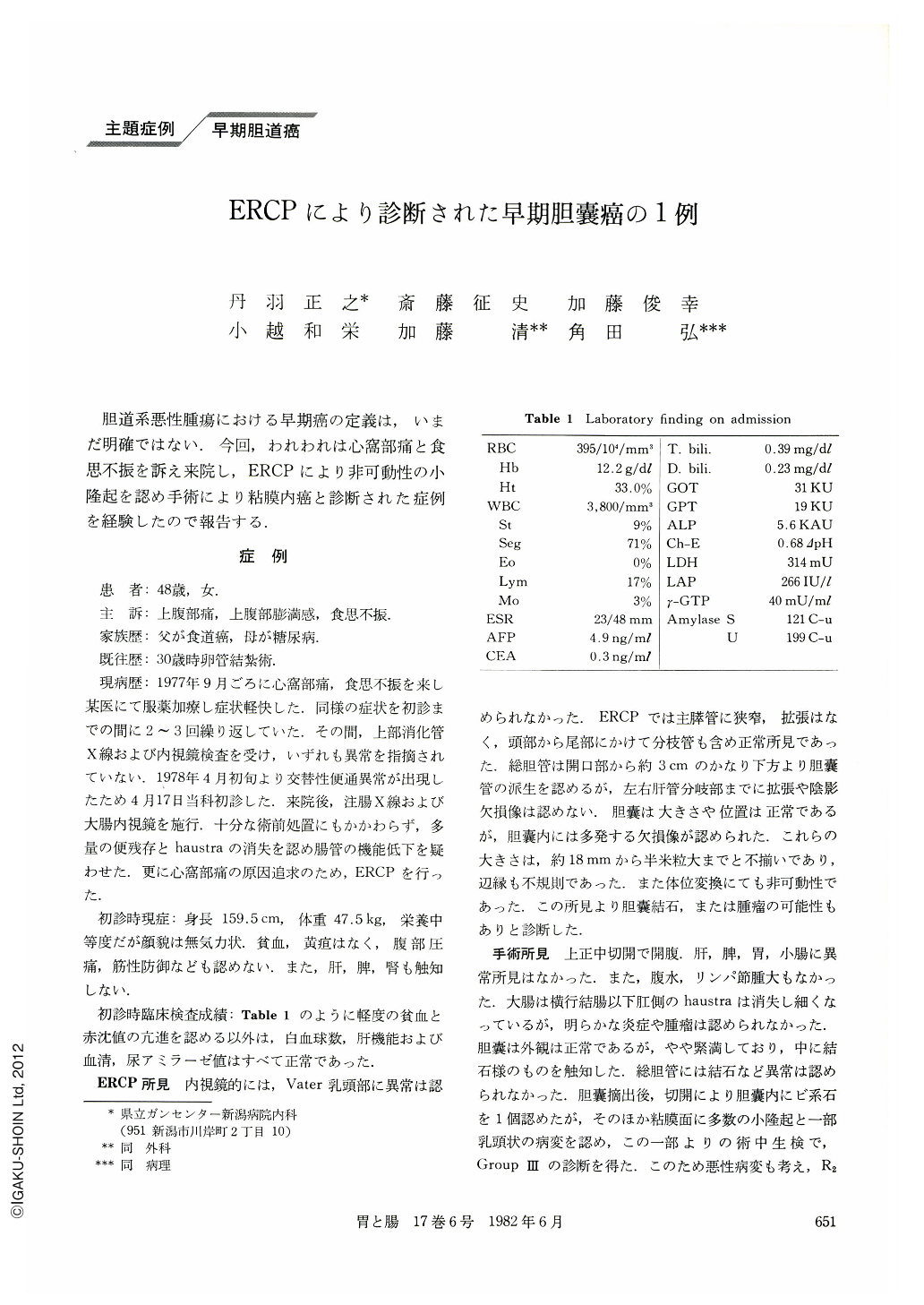
The initial laboratory examination showed only slight anemia and accelerated BSR. Liver function tests were normal as were the amylase levels of the blood and urine. As the upper GI series, including endoscopy, were normal, we performed ERCP as a screening. Endoscopically, the papilla of Vater was normal. Pancreatography showed a normal main duct and its branches. The cystic duct branched off from the choledochus at a level of about 3 cm from the distal end of the choledochus. No abnormality was seen up to the intrahepatic ducts. In the gallbladder multiple shadow defects of various sizes were seen, ranging from half the size of a rice grain to about 18 mm in diameter. Their margins were irregular. Shifts of body positions showed that they were unmovable, so that we suspected gallbladder tumors (polyps) or intramural gallstones. Surgery disclosed a single gallstone of bilirubin origin within the gallbladder and several Ⅱa-like small protrusions on the mucosal surface. Also a papillary fleck was seen within the gallbladder, 20×15×10 mm in size, which was partly isolated. As a tissue analysis of this specimen during the operation showed Group Ⅲ, malignant changes were also considered. A complete resection of regional lymph nodes (R2) was performed.
Pathological findings of the excised gallbladder showed that the isolated papillary fleck was a part of a polypoid lesion which had fallen from the body of the gallbladder. The diagnosis was papillary adenocarcinoma. The same change was also seen at the base, but the depth of the invasion was confined to the mucosa. As no metastasis was seen in the resected lymph nodes, we arrived at a diagnosis of early cancer of the gallbladder.
Copyright © 1982, Igaku-Shoin Ltd. All rights reserved.