Japanese
English
- 有料閲覧
- Abstract 文献概要
- 1ページ目 Look Inside
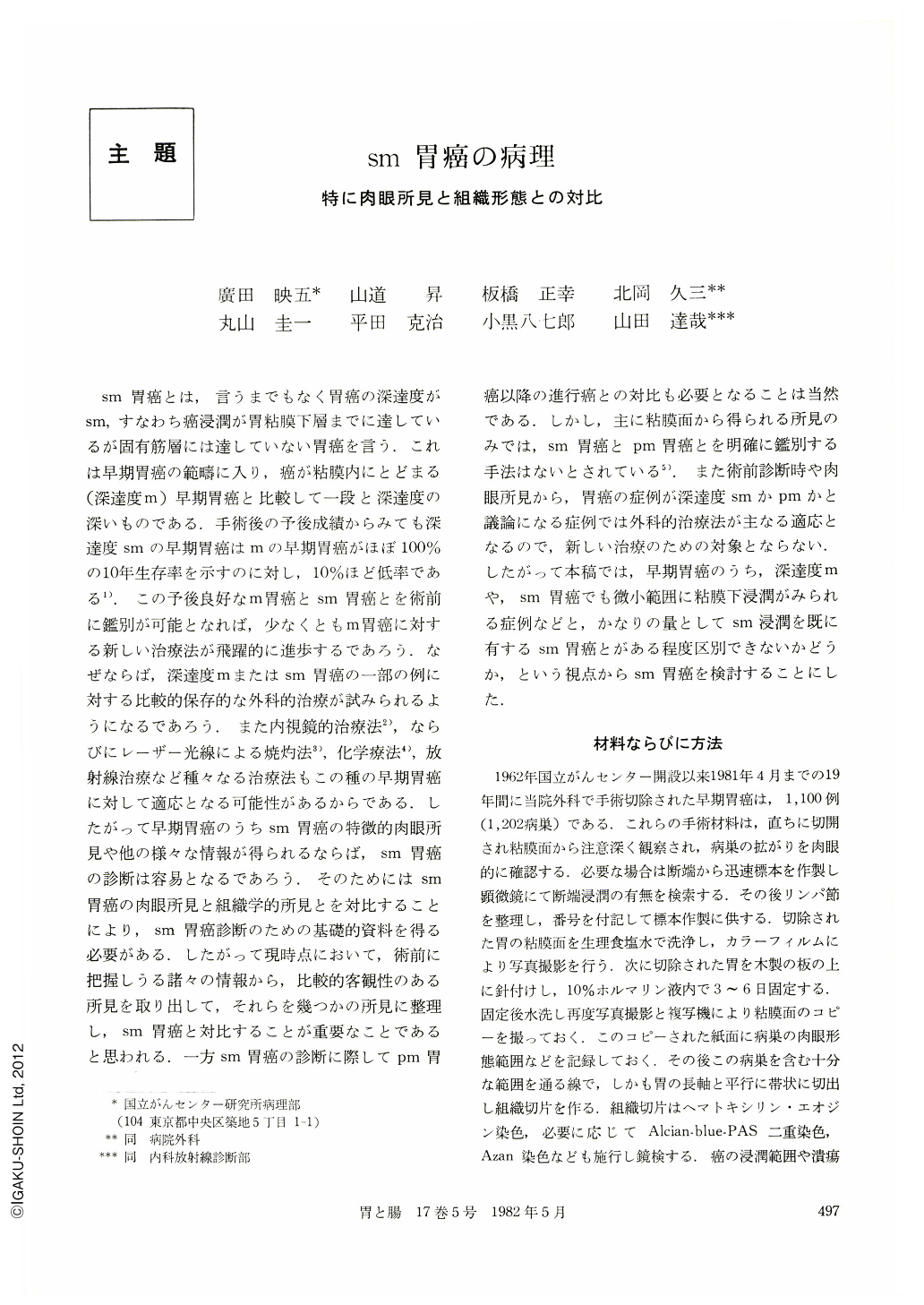
sm胃癌とは,言うまでもなく胃癌の深達度がsm,すなわち癌浸潤が胃粘膜下層までに達しているが固有筋層には達していない胃癌を言う.これは早期胃癌の範疇に入り,癌が粘膜内にとどまる(深達度m)早期胃癌と比較して一段と深達度の深いものである.手術後の予後成績からみても深達度smの早期胃癌はmの早期胃癌がほぼ100%の10年生存率を示すのに対し,10%ほど低率である1).この予後良好なm胃癌とsm胃癌とを術前に鑑別が可能となれば,少なくともm胃癌に対する新しい治療法が飛躍的に進歩するであろう.なぜならば,深達度mまたはsm胃癌の一部の例に対する比較的保存的な外科的治療が試みられるようになるであろう.また内視鏡的治療法2),ならびにレーザー光線による焼灼法3),化学療法4),放射線治療など種々なる治療法もこの種の早期胃癌に対して適応となる可能性があるからである.したがって早期胃癌のうちsm胃癌の特徴的肉眼所見や他の様々な情報が得られるならば,sm胃癌の診断は容易となるであろう.そのためにはsm胃癌の肉眼所見と組織学的所見とを対比することにより,sm胃癌診断のための基礎的資料を得る必要がある.したがって現時点において,術前に把握しうる諸々の情報から,比較的客観性のある所見を取り出して,それらを幾つかの所見に整理し,sm胃癌と対比することが重要なことであると思われる.一方sm胃癌の診断に際してpm胃癌以降の進行癌との対比も必要となることは当然である.しかし,主に粘膜面から得られる所見のみでは,sm胃癌とpm胃癌とを明確に鑑別する手法はないとされている5).また術前診断時や肉眼所見から,胃癌の症例が深達度smかpmかと議論になる症例では外科的治療法が主なる適応となるので,新しい治療のための対象とならない.したがって本稿では,早期胃癌のうち,深達度mや,sm胃癌でも微小範囲に粘膜下浸潤がみられる症例などと,かなりの量としてsm浸潤を既に有するsm胃癌とがある程度区別できないかどうか,という視点からsm胃癌を検討することにした.
This study was carried out in order to make it clear what kind of macroscopic and histologic findings obtained preoperatively are the most important to diagnose submucosal invasion of gastric cancer. One thousand and a hundred cases (1,202 cancerous lesion) of early gastric cancer were used for this study, which received surgical resection at National Cancer Center Hospital during the period from May 1962 to April 1981. With these materials, we examined clinicopathologically the frequency and mode of submucosal invasion of cancer and their relation with several possibly important macroscopic and microscopic findings. The results obtained are as follows.
(1) Age and Sex: Peak of age distribution of gastric cancer with submucosal invasion (sm cancer) was at the seventh decade and average age was 56.7 years, which were almost same as those of whole (intramucosal and submucosal) early gastric cancer cases. Male-female ratio was 2.53. Incidence of sm cancer increased with aging in male group but not in female group.
(2) Localization: Occurence of sm cancer was found most frequently in the M-area along the lesser curvature (25.5%), which was, however, lower than that of whole early gastric cancer. It was relatively higher in both anterior and posterior wall of M-area in comparison with those of the whole early gastric cancer.
(3) Gross types: Common gross types of sm cancer were Ⅱc type (53.1%), Ⅱa+Ⅱc type (12.3%) and Ⅰ type (11.2%). Ⅱa+Ⅱc type early gastric cancer showed the highest frequency of submucosal invasion (63.1%).
(4) Histologic types: Differentiated type and undifferentiated type adenocarcinoma were almost equal in the frequency of sm cancer. Poorly differentiated adenocarcinoma showed submucosal invasion more frequently (56.9%) than the other histological types except mucinous one.
(5) Association of peptic ulcer or ulcer scar within the cancerous lesion: It was found in 68% of sm cancer cases. Association of ulcer or ulcer scar increased the frequency of submucosal invasion in Ⅱc type(46.4%), compared with 26.7% of submucosal invasion in non-ulcerative Ⅱc type. Poorly differentiated adenocarcinoma showed higher frequency of submucosal invasion in ulcerative group(60.5%)than in non-ulcerative one (44.7%).
(6) Size of the cancerous lesion: Submucosal invasion was present in 17.4% of early gastric cancer smaller than 1 cm in diameter and increasted in the frequency as the size of the lesion becomes larger (2~5cm: sm 40~50%, 5cm: sm 50~60%). Non-ulcerative depressed type cancer smaller than 1 cm showed very low frequency of submucosal invasion. Ⅱa+Ⅱc type cancer showed submucosal invasion in more than half of the cases even though smaller than 2 cm in the diameter.
(7) Mode of submucosal invasion: It was classified into 4 groups; 1 minute, 2 scattered, 3 expansive and 4 diffuse infiltrative. Most frequent group was “scattered” (44%) and the second “minute” (40%).
(8) Nodal status: Nodal metastasis was found in 13.9% of the whole sm cancer cases. In relation to association of ulcer, sm cancer without ulceration showed higher frequency of nodal metastasis (23.2%) than those with ulcer (9.8%). Intramucosal carcinoma without association of ulcer showed no metastasis to the lymph nodes.
These results may lead to the conclusion that we can estimate the presence and the mode of submucosal invasion of gastric cancer with considerable reliability if we could obtain preoperatively the clinical, macroscopic and microscopic information written above. Necessity of concept of minute gastric cancer for modified treatment was discussed and emphasized in this report.

Copyright © 1982, Igaku-Shoin Ltd. All rights reserved.


