Japanese
English
- 有料閲覧
- Abstract 文献概要
- 1ページ目 Look Inside
大腸ポリープに関するMorson1),武藤2)らの組織学的分類は,その診断および治療の際に極めて有用で,現在広く採用されている.しかしながら,この分類のいかなる範疇にも該当しない症例がまれに存在するようである.われわれは,直腸炎罹患後約6年を経過して発見され,著明な低蛋白血症を伴い,巨大でしかも興味深い組織像を呈した直腸腺腫の1例を経験したので報告する.
We herein report a case of a large adenoma of the rectum associated with marked hypoproteinemia. This adenoma was pointed out about six years after proctitis and this pathology showed some interesting findings.
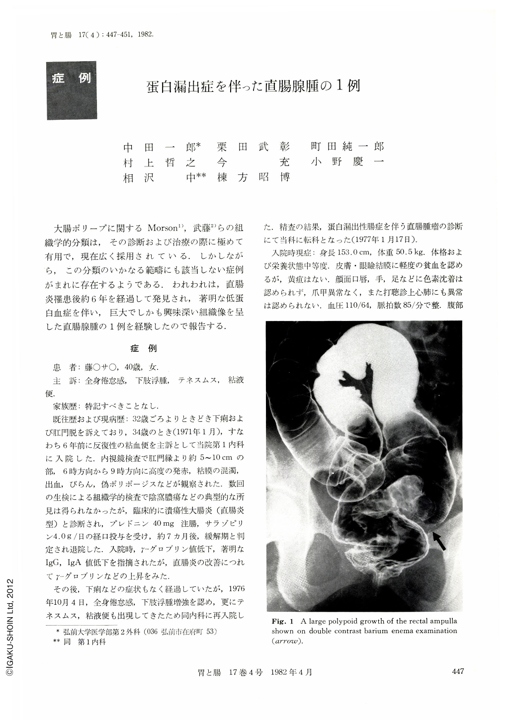
A 40-year-old housewife was admitted to our hospital with general fatigue, edema of the lower extremities, tenesmus and mucus in her stools on October 4, 1976. There was no family history of a remarkable illness. The skin and mucous membrane contained no abnormal pigmentation on physical examination. Digital and proctoscopic examinations revealed a large, lobulated, reddish tumor mass, the lower margin of it being palpable 8 cm proximal to the anal verge. A biopsy of the mass was reported as adenoma. A barium enema demonstrated the presence of a large filling defect in the rectal ampulla, supporting the diagnosis of a rectal tumor (Fig. 1). The other x-ray examination of the stomach, small bowel and colon showed no abnormality. Significant laboratory data were: serum protein value 3. 9 g/dl; γ-globulin 1.1%; IgG 442 mg/dl (normal 800~1400); IgA 46 (150~400). Urinalyses tested negative for albumin on several occasions. The patient had excessive protein loss into the gastrointestinal tract as demonstrated by 131I-PVP test 6.4% (normal 0.5%) (Table 1).
She was treated by per-anal local excision with the diagnosis of rectal adenoma associated with protein-losing enteropathy (Fig. 2). Macroscopically, the rectal tumor obtained was lobular, reddish, here and there coated, 12×3.0×2.5 cm in size and 77 grams in weight (Fig. 3). Microscopically, it was composed of an overgrowth and elongation of the colonic epithelium which was slightly dysplastic (Fig. 4). The surface epithelium had almost disappeared. The superficial region of the tumor was covered with a fibrinous exudate and necrotic cells, and was very heavily infiltrated with inflammatory cells. Dilatated lymphatic channels were not present in mucoua and in submucosa (Fig. 5). Hypoproteinemia improved after the excision of the rectal tumor (Table 2). However, about nine months later the same lesions occurred again. A low anterior resection of the rectum was performed (Fig. 6 a, b).
We consider that this rectal lesion may not come under every category of the common histological classification of polyps of the large intestine.
Copyright © 1982, Igaku-Shoin Ltd. All rights reserved.