Japanese
English
- 有料閲覧
- Abstract 文献概要
- 1ページ目 Look Inside
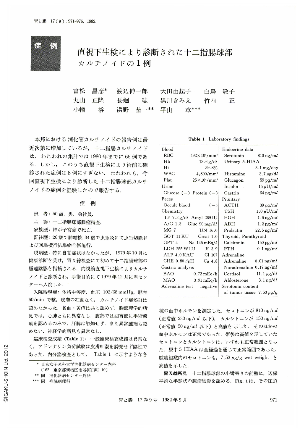
本邦における消化管カルチノイドの報告例は最近次第に増加しているが,十二指腸カルチノイドは,われわれの集計では1980年までに66例である.しかし,このうち直視下生検により術前に確診された症例は8例にすぎない.われわれも,今回直視下生検により診断した十二指腸球部カルチノイドの症例を経験したので報告する.
A -50-year-old man received gastric mass screening although he had no symptoms to complain of. At this time he was found to harbor a round tumor in the duodenal bulbus by upper gastrointestinal roentgenograms and it was diagnosed histologically as carcinoid tumor by endoscopic biopsy.
The tumor, measuring 8×7×5 mm in size, was removed by surgical operation. It revealed a hemispheric shape and its cut surface was yellow. Microscopic examination revealed a carcinoid composed of tumor cells of uniform size with trabecular structure. Argyrophil reaction by Grimelius method showed argyrophil granules, but the argentaffin reaction by Fontana-Masson method was negative.
Electromicroscopic finding revealed dense and round secretory granules, measuring 200~300 nm, in the cytoplasm of the tumor cell.
Serum serotonin and calcitonin were elevated above normal values in the preoperative measurement, but urinary content of 5-HIAA was within normal level. After operation, serum serotonin and calcitonin returned to normal. The carcinoid tumor was found to contain 7.53 μg of serotonin pergram wet weight of tissue.
So far we have been able to collect 67 reported cases of duodenal carcinoid in Japan including the present case, but only nine of them had been diagnosed by endoscopic biopsy before operation.
Copyright © 1982, Igaku-Shoin Ltd. All rights reserved.