Japanese
English
- 有料閲覧
- Abstract 文献概要
- 1ページ目 Look Inside
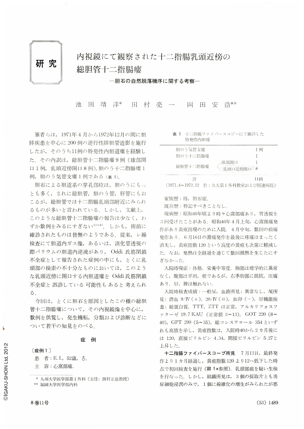
筆者らは,1971年4月から1972年12月の間に胆膵疾患を中心に200例の逆行性膵胆管造影を施行したが,そのうち11例の特発性内胆道瘻を経験した.その内訳は,総胆管十二指腸瘻9例(球部開口1例,乳頭近傍開口8例),胆のう十二指腸瘻1例,胆のう気管支瘻1例である(表1).
胆石による胆道系の穿孔部位は,胆のうにもっとも多く,まれに総胆管,胆のう管,肝管にもおこるが,総胆管では十二指腸乳頭部附近にみられるものが多いと言われている.しかし,文献上,このような総胆管十二指腸瘻の報告は少なく,わずか数例をみるにすぎない1)~3).しかも,術前に確診されたものは皆無のようである.従来,レ線検査にて胆道内ガス像,あるいは,消化管透視の際バリウムの胆道内逆流があり,Oddi氏筋閉鎖不全症として報告された症例の中にも,とくに乳頭部の検索の不十分なものにおいては,このような乳頭近傍に開口する内胆道瘻をOddi氏筋閉鎖不全症と誤診している可能性もあると考えられる.
During the period April 1971 to Dec. 1972, we have performed endoscopic pancreatocholangiography centering on patients with diseases of the pancreas and biliary tract. This paper deals with 8 cases we have encountered of choledochoduodenal fistulae in the neighborhood of the duodenal papilla, suggesting natural evacuation of gallstones. Emphasis is laid on their endoscopic pictures.
Ⅰ. Choledochoduodenal fistulae opening into neighborhood of the papilla were divided into two types according to their size and location. Type type Ⅰ (Cases 1, 2, 3 and 4): The fistulae were small and opened into oral part adjoining the papilla orifice, or above the so-called longitudinal fold. Anatomically, they were located in the intramural duct. Type Ⅱ (Cases 5, 6 and 7): The fistulae were larger and away from the papilla orifice, adjacent the oral part of the longitudinal fold. Their anatomical site was in the common bile duct just before the intramural bile duct.
Ⅱ. X-ray findings: Gas or barium was not observed in the biliary tract in type Ⅰ, while in type Ⅱ pneumobilia was recognized, with the contrast medium easily flowing up into the biliary tract during fluoro-scopy of the digestive tract.
Ⅲ. Pathogenesis: The 8th case with fistula and impacted gallstone we were able to observe endoscopically attests to the formation of the internal biliary fistula by compression of the stone on the biliary lumen and its subsequent necrosis. The difference between the type Ⅰ and Ⅱ was regarded as owing to the difference in size of the gallstones. In other words, type Ⅰ fistula was caused by gallstones small enough to be impacted in the intramural bile duct, while that of type Ⅱ resulted from stones too large to be impacted there.
Ⅳ. It has been demonstrated that, besides incontinence of the sphincter of Oddi, some cases of choledochoduodenal fistula of type Ⅱ are responsible for the regurgitation of the contrast medium up into the biliary tract.
Copyright © 1973, Igaku-Shoin Ltd. All rights reserved.