Japanese
English
- 有料閲覧
- Abstract 文献概要
- 1ページ目 Look Inside
- サイト内被引用 Cited by
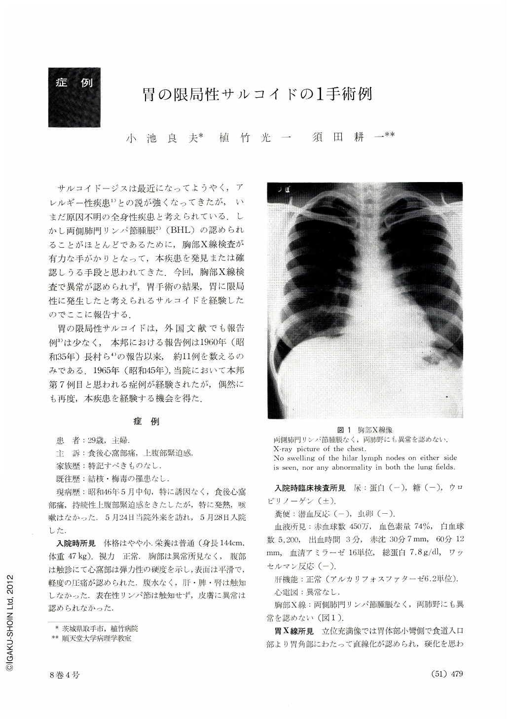
サルコイドージスは最近になってようやく,アレルギー性疾患1)との説が強くなってきたが,いまだ原因不明の全身性疾患と考えられている.しかし両側肺門リンパ節腫脹2)(BHL)の認められることがほとんどであるために,胸部X線検査が有力な手がかりとなって,本疾患を発見または確認しうる手段と思われてきた.今回,胸部X線検査で異常が認められず,胃手術の結果,胃に限局性に発生したと考えられるサルコイドを経験したのでここに報告する.
胃の限局性サルコイドは,外国文献でも報告例3)は少なく,本邦における報告例は1960年(昭和35年)長村ら4)の報告以来,約11例を数えるのみである.1965年(昭和45年),当院において本邦第7例目と思われる症例が経験されたが,偶然にも再度,本疾患を経験する機会を得た.
The patient, a 29-year-old housewife, visited our clinic complaining chiefly of a pain in the epigastric region after meals and continuos strain in the upper abdomen. On palpation, the epigastric region showed a hardnesss of an elastic nature, had an even and smooth surface and was slightly tender to pressure. Roentgenogram of the chest showed no swelling of the hilar lymph nodes on either side (BHL) nor any abnormality in the lung fields. After roentgenographic and endoscopic observation, the case was diagnosed as scirrhous cancer of the stomach, and total gastrectomy was performed. The whole wall of the extirpated stomach was thickned in an edematous manner. On the surface of the mucosa, there were comparatively superficial ulcerations of irregular shape centering around the gastric body. Palpation and visual examination gave an impression of “soft scirrhous cancer”.
As the result of histopathological examinations, granulomas consisting of epithelioid cells along with Langhans' giant cells were found distributed extending over both the proper layers of the mucosa and the serous membranes. Henes the case was considered gastric sarcoid. Tests for Kveim's and tuberculin reactions conducted postoperatively were both negative. At present, as of November 1972, one year and four months after operation, the patient is doing well, showing no abnormality worthy of special mention.
Our patient is considered the twelfth case in our country of localized gastric sarcoid. Ten out of twelve patients were women, and they chiefly complained of a pain in the epigastric region or/and the upper abdomen. The diagnoses were, in all the cases, established by postoperative histological examination. They are broadly classified into 2 groups: the one, after gastrectomy for stomach ulcer, was accidentally found to be sarcoid by histological examination, and the other had been suspected of scirrhous cancer of the stomach but did not prove so histologically. Furthermore, the fact that X-ray examination of the chest showed no BHL in any of the cases is one of the points worthy of consideration in actually making clinical examination.
Copyright © 1973, Igaku-Shoin Ltd. All rights reserved.