Japanese
English
- 有料閲覧
- Abstract 文献概要
- 1ページ目 Look Inside
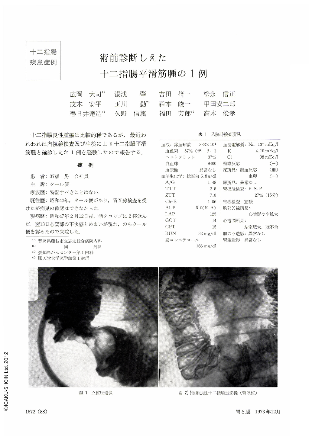
十二指腸良性腫瘍は比較的稀であるが,最近われわれは内視鏡検査及び生検により十二指腸平滑筋腫と確診しえた1例を経験したので報告する.
This is a report of a leiomyoma of the duodenum, relatively a rare occurrence, accurately diagnosed as such by endoscopy and biopsy. The patient, a 37-year-old man with a chief complaint of tarry stool, had previously in 1967 experienced a bout of it. On February 12, 1972, he had another episode of tarry stool and was admitted to the hospital. Upper GI x-ray series disclosed a round shadow defect, measuring 44 mm in diameter, at the junction between the second and third limb of the duodenum. Duodenofiberscopy with JFB disclosed in the abovementioned site a hemispheric tumor, its surface looking norma and accompanied with bridging folds. There was a deep ulcer in the center under a diagnosis of submucosal tumor of the duodenum we took out several pieces of specimens under direct vision from the ulcer floor. Our diagnosis then was leiomyoma of the duodenum. Surgical intervention showed a yamshaped tumor, 40×50×35 mm, developing mostly outside the duodenal wall. In the center was an ulceration, measuring 11×5 mm. Histological findings were those of leiomyoma almost the same as had been obtained at biopsy. Diagnostic principles of x-ray and endoscopy for submucosal tumors of the stomach can also be applied to those of the doudenum, but their accurate diagnosis is difficult, generally speaking. When such a tumor is associated with ulceration, tumor tissue can be extracted under direct vision.
Copyright © 1973, Igaku-Shoin Ltd. All rights reserved.