Japanese
English
- 有料閲覧
- Abstract 文献概要
- 1ページ目 Look Inside
腸結核は,現在でも稀な病変ではない.偶然に発見される腸結核もめずらしくない.最近ではクローン病との鑑別において注目されている.今回,胆石をもち,右半結腸に病変を認め腸結核と思われる1例を経験したので報告する.
A 57-year-old female has had diarrhea and abdominal discomfort since about 35 years old. She was admitted to our hospital on October in 1977 after daily diarrhea of ten years’ duration. She had a mild anemia. Chest X-ray showed a small calcified lesion in both sides of pulmonary hilus. Cho1ecystography and ERCP disclosed numerous calculus in gall bladder.
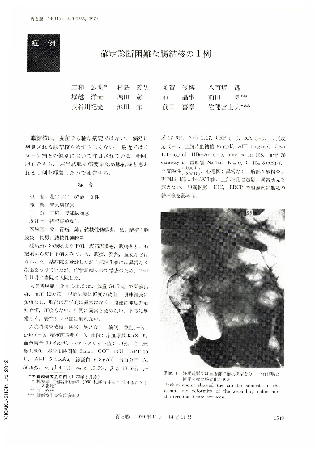
Roentgengraphic examination: Double contrast study of the colon showed disappearance of colonic haustration with rough mucosal surface from anal to one third of transverse colon. Cecum seemed to be disappeared and ascending colon merged into ileum directly, but there were apparent stricture at ileo-cecal region.
The double contrast radiograph with a moderate volume air showed linear ulcer scars running circulary to the stenotic region, fissuring ulcer at both sides of the wall and pseudo-diverticular lesions at the proximal as well as distal portion of the stenosis. It also showed mucosal bridges between ulcer scars and various sizes of pseudopolys. A large volume air radiograph showed the prominent pseudopolyps.
Endoscopic examination: No ulcers of active stage were detected and no granuloma could be found by biopsy.
Pathological findings: Cholecystectomy and hemicolectomy was performed. The resected colon specimen showed circular linear ulcer scar forming stricture at ileo-cecal region. There were also scattered multiple bridges and pseudopolyps around the stricture.
In the histological study, ulcer scars were mostly noted as U1-Ⅱ. But partially it was found that the stages of ulcer were Ul-Ⅲ or Ul-Ⅳin both ends of mucosal bridge. But granuloma was not found at all. Hyalinization or paramyloid were noted only in a part of the lymph node, and tubercle bacillus as well as caseous granuloma were not detected. By the X-ray findings and reference of literature, our case was diagnosed as tuberculosis of the intestine.
Copyright © 1979, Igaku-Shoin Ltd. All rights reserved.