Japanese
English
- 有料閲覧
- Abstract 文献概要
- 1ページ目 Look Inside
虚血性大腸炎は血管造影で異常を示すことは少ない.われわれは血管造影で診断された上腸間膜動脈病変,動脈瘤,腸間膜血腫を伴う特異な1例を報告する.
症 例
患 者:52歳 男
主 訴:下腹部仙痛
家族歴,既往歴に特記すべきことはない.
現病歴:1977年12月9日午後7時頃,自宅で,突然下腹部に仙痛を覚えた.安静臥床によっても軽減せず,同日午後10時にいわき市立常盤病院を受診した.
初診時,血圧,脈拍,呼吸は正常であったが,顔貌は苦悶状であった.黄疸,貧血の微候もなく,胸部,肺野に所見なく,心雑音も聴取されず,腹部では肝・脾・腎も触れなかった.嘔吐,下痢,血便などはなかったが,胸部から下腹部にかけてかなり激しい疼痛を訴えたので急性腹症として即日入院した.
A 52-year-old man was well until December 9, 1977, when he suddenly developed colic in the evening and visited the hospital because the pain persisted.
Physical examination revealed he was acutely ill, but no other objective findings were elicited except abdominal tenderness. He was admitted as acute abdomen. On the following day, he again developed colic in the right upper quadrant and fell into shock in the evening. He recovered from shock within 30 minutes and became ambulatory as usual on the next day.
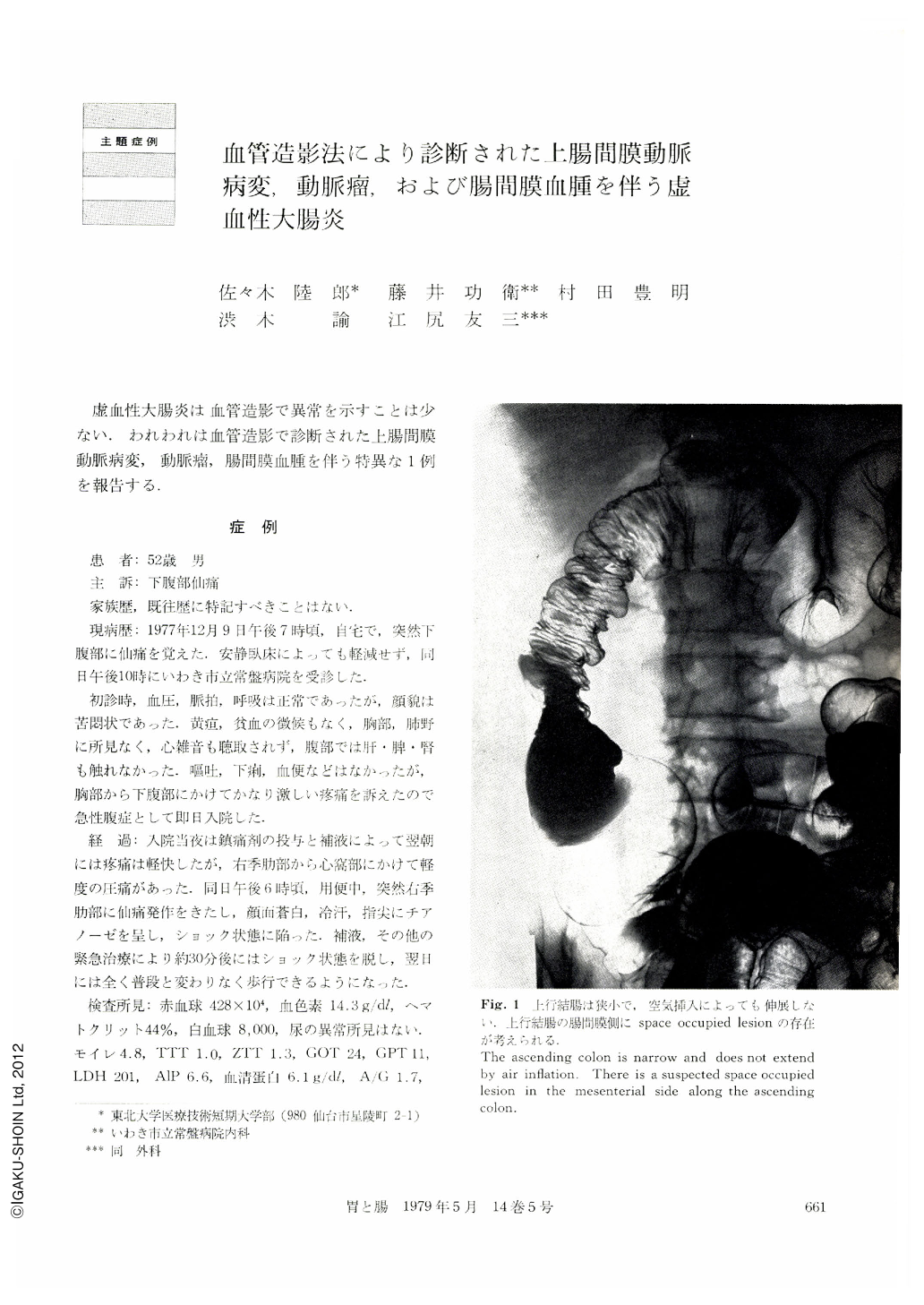
Routine laboratory studies revealed no abnormalities. Plain film of the abdomen, upper GI series and cholecystogram were also non-revealing. However, barium enema demonstrated a narrow portion of the ascending colon and probably a space occupied lesion in the mesenterial side along the ascending colon. Selective angiogram of the superior mesenteric artery demonstrated a narrow trunk a few cm from the orifice to the origin of the ileocolic artery and complete occlusion of the middle colic artery and right colic artery. There were two aneurysms at the trunk of the superior mesenteric artery. Segmental absence of vasa recti in the ascending colon was seen. In addition, a space occupied lesion was suspected in the mesenterial side along the ascending colon.
From these findings, the diagnosis of ischemic colitis associated with a narrow trunk and aneurysms of the superior mesenteric artery and mesenteric hematoma possibly due to rupture of an aneurysm, was made, and laparotomy was performed on December 27, 1977.
Findings at operation were as follow: 1) Pulsations were not felt on the middle colic artery and the right colic artery, though there was a weak pulsation on the stenotic superior mesenteric artery. 2) There were two small aneurysms at the superior mesenteric artery. 3) The segmental portion of the middle ascending colon was definitely narrow but the appea-rance was normal. 4) There was a tumor with size of 9×6×3 cm in the mesenterial side along the ascending colon.
Microangiography of the resected specimen demonstrated absence of vasa recti in the middle portion of the ascending colon. Resected specimen showed a definite color change and narrow hollow viscus, indicating ischemia in the ascending colon. The cut surface of the mesenterial tumor showed a fairly fresh hematoma.
It has been reported that ischemic colitis occurs predominantly in the left side colon and vascular lesions are not always demonstrated on angiographic examinations. Since the reported case had ischemic lesion in the right side colon, the narrow trunk of superior mesenteric artery, obstruction of the middle colic artery and right colic artery and two small aneurysms, angitis rather than atherosclerotic changes is most likely. Mesenteric hematoma was thought to be due to rupture of a small aneurysm, when considering clinical course. From these reasons, this case was considered to be a rather unusual case of ischemic colitis.
Copyright © 1979, Igaku-Shoin Ltd. All rights reserved.