Japanese
English
- 有料閲覧
- Abstract 文献概要
- 1ページ目 Look Inside
Computerised Tomography(CT)はX線を診断の目的で使用する極めて有効な検査法である.従来のX線検査では認められなかった組織間の細かいX線吸収差が表現され,まず脳疾患X線診断に応用されて驚異的な成功を収めた(Ambrose,1973,Paxton & Ambrose,1974).この装置の発明者G.N. Hounsfieldの業績の偉大さは医療に従事するものがいかにCTを高く評価しているかによって明らかである.脳疾患は侵襲を加えることなしに描出され,病変の早期診断が可能で,重篤な患者でも状態を悪化させることなく検査が可能である.CTが利用できる脳神経放射線科では気脳検査と血管造影の件数が著明に減少した(Ambrose et al,1976).
脳疾患に対するCTの疑いない診断価値は他の身体部分を検査できる装置の開発を促した.全身CTの経験は現在急速に集積されているが重要なことは全身CTで脳疾患も診断できることである.全身CTで得られる脳疾患の画像は最新の頭部専用CTの画像と比肩できるもので,両機種とも被検者を水でかこむ(water bagの使用)必要はなくなった.
The E.M.I. general purpose scanner produces C.T. scans of the brain and base of the skull as well as the rest of the body. The brain pictures are comparable in quality to that of the latest head equipment and the bones and soft tissue of the base of the skull are shown in great detail. It is therefore particularly valuable for showing soft tissue space occupying lesions and osteolytic and sclerotic deposits in and around the base of the skull in the investigation of proptosis, glomus jugulare tumours and carcinoma of the nasopharynx.
C.T. has been found to be particularly valuable in showing the extent of malignant disease and in lymphoma and testicular tumours. Lymph node enlargement and tumour recurrence in the abdomen, thorax and axillae can be outlined when not demonstrated by other radiological methods including conventional tomography, lymphography and arteriography and the information has proved invaluable for radiotherapy, chemotherapy or surgery in these patients.
Unusual and unsuspected aspects of disease have been shown such as the not uncommon finding of early pleural effusions, pulmonary oedema and ascites and rarely the presence of sarcoma metastases in muscle. Even the physiological changes in the size of peripheral pulmonary artery branches with change in position from supine to prone can be demonstrated.
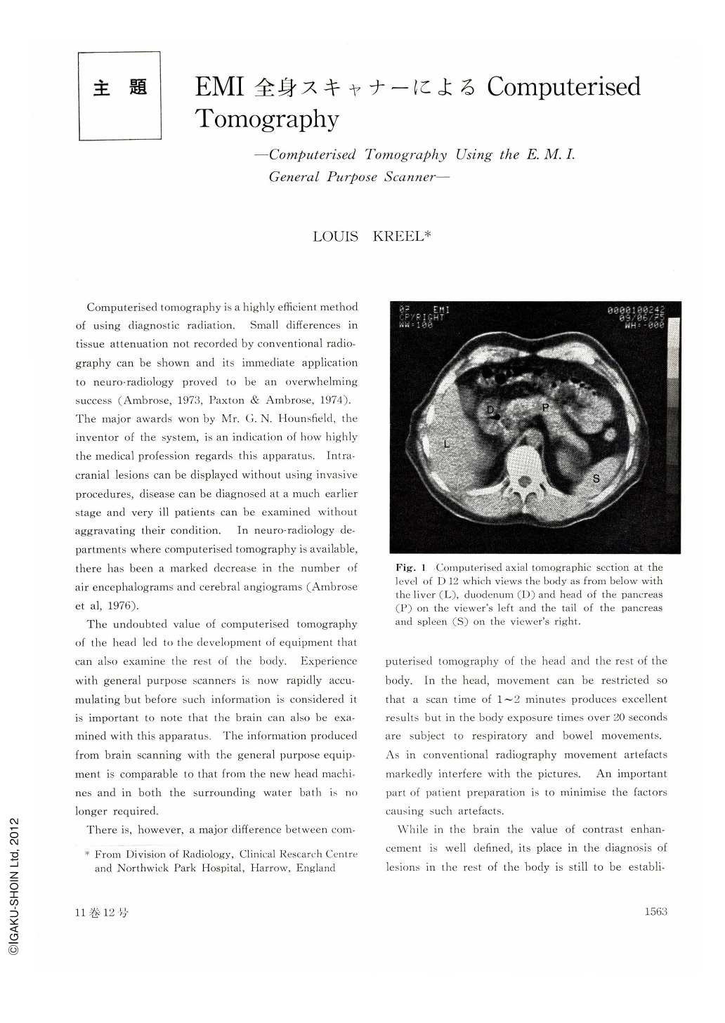
Retroperitoneal tumours, mass lesions of the pancreas, liver and kidneys and surrounding tissue infiltration show quite clearly. Cysts and abscesses can be accurately differentiated from solid lesions and associated bone involvement, whether by direct spread or by metastases.
The delineation of tumours in axial section lends itself to accurate radiotherapy planning and monitoring of the effects of treatment. This information is similarly valuable in chemotherapy. The display of tumour recurrence allows surgeons more effective management of these cases especially in the decision as to where and when to operate.
It is hoped that future developments will allow accurate display of the spinal cord, intervertebral disc material and of the heart. Volumetric studies of cardiac chambers and the display of normal and abnormal myocardium could well prove extremely important in the management of patients with angina, myocardial infarction and ventricular septal defects.
While there is already conclusive evidence of the assured place of computerised tomography in neuroradiology its place in whole body scanning is yet to be established. However, it would appear that on present evidence a similar position holds at least with malignant disease in diagnosis and the assessment of the effects of treatment. This applies particularly to lymphoma, testicular tumours and retroperitoneal tumours.
Copyright © 1976, Igaku-Shoin Ltd. All rights reserved.