Japanese
English
- 有料閲覧
- Abstract 文献概要
- 1ページ目 Look Inside
- サイト内被引用 Cited by
胃癌の1つであるいわゆるスキルスは,一般的に,胃癌の中で予後の悪い癌であるとされている.そして,わが国においてはスキルスといった場合には,癌の肉眼型であるBorrmann 4型あるいはlinitis plasticaを意味する場合がある1)12)26)27)30).一方では組織水準で,スキルスは胃癌の特徴ある組織型の1つとして硬癌adenocarcinoma scirrhosum22),scirrhous carcinoma13),carcinoma fibrosum3)7)と呼ばれている.さらには,このような組織型は乳癌にも多いものである.このように,いわゆるスキルスという言葉は胃においては肉眼的水準および組織学的水準での両方の意味を含んでいて混乱を与えている.スキルスという言葉を用いる場合にはどの臓器でどの水準の意味で用いているのかを明確にしておく必要がある.
著者らは,スキルスという言葉は癌の形態ではなく癌の質的なことを意味するものであるとの立揚から,胃におけるスキルスの臨床的ならびに病理組織学的なことについて統一的にながめてみた.
The purpose of the present paper is to summarize clinical and histopathological features of scirrhous carcinoma of the stomach which is synonymous with carcinoma fibrosum (Borrmann, R.; Konjetzny, G. E.), diffuse carcinoma (Järvi, O. H., Lauren, P.), diffusely infiltrative carcinoma (Ming, Si-Ch.), and undifferentiated adenocarcinoma (Nakamura, K.), and furthermore to deduce a period of time from cancer development to the leather bottle stomach.
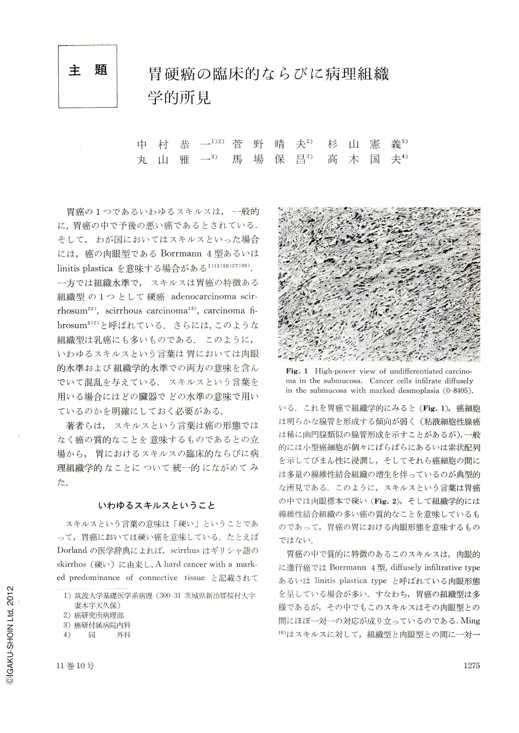
Histologically, cells of scirrhous carcinoma are small and scatteringly present in dense fibrous tissue (Fig. 1), and are rare to form glandular pattern. This typical pattern of scirrhous carcinoma is recognized in the gastric layers except the mucosa. In the mucosa involved by scirrhous carcinoma cells, there is no proliferation of connective tissue. In the mucosa, cells of scirrhous carcinoma diffusely infiltrate without formation of gland, or show signet ring cell type, or show trabecular arrangement (Fig. 6). There is nearly one-to-one correspondence between these histological patterns in the mucosa and scirrhous carcinoma. Consequently, it is evident that intramucosal carcinoma showing these three patterns is early phase of scirrhous carcinoma. Carcinoma showing these three patterns in the mucosa and scirrhous carcinoma belong histogenetically to the same category classified as undifferentiated adenocarcinoma. The undifferentiated adenocarcinoma has been named from the view-point of structure atypism, and is a general term for these three histological patterns in the mucosa and scirrhous carcinoma in the gastric layers except the mucosa. The undifferentiated adenocarcinoma arises from the ordinary mucosa of the stomach consisting of the pyloric, fundic and cardiac gland mucosae, and is characteristic of diffuse infiltration in the gastric wall.
Macroscopically, the majority of undifferentiated adenocarcinoma shows depressed type (IIc, IIc+III, III+IIc) in early phase, and shows diffusely infiltrative type (Borrmann 4, diffuse carcinoma, diffusely infiltrative carcinoma) in advanced phase (Fig. 3).
The undifferentiated adenocarcinoma becomes infrequently a state called linitis plastica type. As to primary location of carcinoma of linitis plastica type, the undifferentiated carcinoma having arisen from the fundic gland mucosa (the corpus) becomes more frequently linitis plastica than the one from the pyloric gland mucosa (the antrum) (Fig. 4). This reason may be that it is clinically difficult to disclose a small lesion in the corpus of the stomach lined with the fundic gland mucosa except the lesser curvature site. Therefore, the undifferentiated adenocarcinoma arising from the fundic gland mucosa is shown to be linitis plastica in many instances.
It has been deduced that a period of time from cancer development to linitis plastica (leather bottle stomach) is several years (about 6 years) (Table 4).
The prognosis of patients having the undifferentiated adenocarcinoma has been generally interpreted as worse than that of patients having differentiated carcinoma, such as tubular or papillary adenocarcinoma. However, the prognosis of patients having the undifferentiated adenocarcinoma measuring less than 4 cm in the largest diameter is better than that of patients having the differentiated adenocarcinoma (Fig. 10). In carcinoma measuring more than 4 cm in the largest diameter and infiltrating up to the proper muscle, the prognosis of patients having the undifferentiated is also better than that of patients having the differentiated (Fig. 11). Meanwhile, in patients operated on for carcinoma penetrating the proper muscle and measuring more than 4 cm in the largest diameter, the prognosis is worse in the undifferentiated than in the differentiated (Fig. 11).
Copyright © 1976, Igaku-Shoin Ltd. All rights reserved.