Japanese
English
- 有料閲覧
- Abstract 文献概要
- 1ページ目 Look Inside
急性上部消化管出血に対しvigorous diagnostic approach1)2)を行なう考えは広く受け入れられた概念となっている.特に内視鏡を用いた早期あるいは緊急検査が出血源診断に安全かつきわめて有用である点を強調した報告3)~10)は多い.
さて本邦においては消化管内視鏡検査が広く普及している反面,出血に対する内視鏡検査には保守的傾向があったものと思われる.しかし最近におけるすぐれた報告11),綜説12),また欧米学者による経験の紹介等13)~15)があり,現在急速に普及しつつあるものと考えられる.
さて,これまでの報告の多くはfiberoptic panendoscopeの開発以前のものであったが,この型の内視鏡の開発により,食道,胃,十二指腸球部および下行脚部の観察は一層能率的かつ合理的となり,すでにpanendoscopeによる緊急内視鏡検査成績16)も報告されている.
By a fiberoptic panendoscope (Olympus GIF-D), emergency endoscopy was performed on 115 patients (60 males, 55 female; mean age-52 years, ranging from 12 to 85 years) where acute upper gastrointestinal hemorrhage was severe enough to necessitate blood transfusion. Soon after resuscitation from shock, endoscopic examination was performed as an initial approach to detect the site of bleeding. The majority (95%) of the patients were endoscoped within 8 to 24 hours after admission to the hospital or initial consultation with the endoscopy team. The remaining 5% of the patients were endoscoped within 24 to 48 hours after recovery from hypotension. Prior to the emergency endoscopy, nasogastric aspiration was performed routinely.
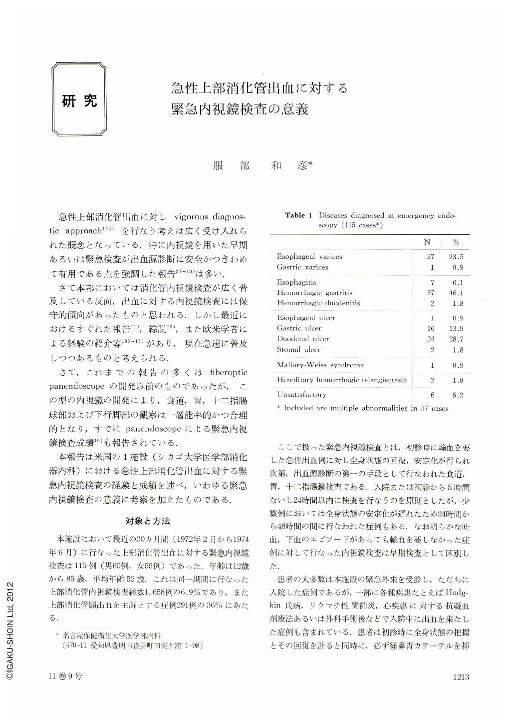
Gastric lavage with ice cold saline or tap water was used when the gastric aspirate contained fresh or coagulated blood. In addition to physical examination, measurement of central venous pressure, ECG, chest and abdominal X-rays, complete blood counts, platelet count, prothrombin time and a screening biochemistry profile were obtained. In these 115 patients, a satisfactory diagnosis was established in 109 cases (95%), while in 6 cases diagnosis was unsatis-factory because of retained blood. A single lesion was detected in 72 patients (63%), while multiple diagnoses were made in 37 patients (32%). Esophagel varices, hemorrhagic gastritis and peptic ulcer diseases were commonly seen (Table 1).
More than half of patients with esophageal varices and hemorrhagic gastritis were diagnosed at the time of emergency endoscopy, while only 12 and 5.4% of the total number of patients with esophagitis and peptic ulcer were diagnosed at the time of emergency endoscopy. The most common source of bleeding in patients with esophageal varices was hemorrhagic gastritis (Table 2), while apparent evidence of variceal tear or rupture was observed in relatively small number of patients. Because hemorrhagic gastritis is so common and it cannot be readily diagnosed by conventional barium study, and because of the multiplicity of co-existing disorders, esophago-gastroduodenoscopy is mandatory in the initial diagnostic approach to acute upper gastrointestinal bleeding.
Copyright © 1976, Igaku-Shoin Ltd. All rights reserved.