Japanese
English
- 有料閲覧
- Abstract 文献概要
- 1ページ目 Look Inside
上部消化管の肉眼病理学的な研究はレ線,内視鏡診断学の進歩とあいまって,微細な粘膜面の変化の観察に至るまで,著しく進歩した.一方,小腸の肉眼病理学的な観察は特殊性炎症や腫瘍性病変については精細な報告があるが,非特異的な変化や1mm前後の微細な変化についての報告は少ない1).この主な理由は剖検では死後直ちに腸管を固定しないかぎり粘膜の自家融解が起こり,微細な病変を観察し得ないからである.小腸ファイバースコープの開発とその実用化への努力は,小腸粘膜の肉眼病理を大いに進歩させるものと思われる.著者は昭和39年以来,岡山大学第一内科剖検例,川崎医大消化器内科剖検例について,死後Rigor mortisに至る以前に腸管内に10%フォルマリン,または2.5%グルタルアルデヒドを注入し充分に空腸全域を固定し,上部消化管粘膜の肉眼病理について検討を続けて来た.本論文では,十二指腸病変を除き,空腸粘膜の肉眼所見について代表例を呈示し報告し,一部,小腸ファイバースコープ(SIF-Type B)について観察し得た内視鏡像を呈示したい.
In recent years, endoscopical techniques of the small intestine have been utilized in the macroscopical and histological diagnosis of the small intestinal disease. From the endoscopical point of view, endoscopical findings should correspond with the anatomic-pathologic findings.
In this report, the author made exact observation of small intestine by studing 34 autopsy materials as a basic study of the small intestinal endoscopy.
Method: The stomach, duodenum and jejunum of fresh cadavers were fixed with 10% Formalin or 2.5% Glutaraldehyde solutions before autopsy by means of gastroduodenal intubation. The anatomy of the small intestine was carefully examined and the findings in each specimen were recorded by means of diagrammatic sketches and color photography. Histological studies (hematoxylin-eosin and PAS stain) and scanning electron microscopic studies in the region of the mucosal changes were examined.
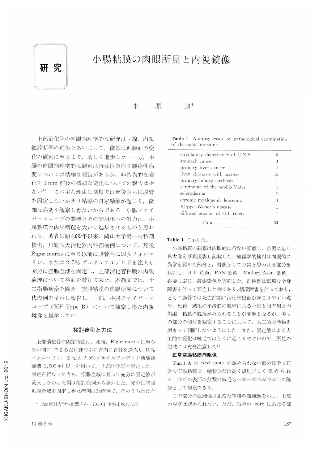
The small intestines of 34 were completely immersed by the fixative without postmortem changes in the morphology of mucosa (Table 1).
Erosion of the small intestine could be a manifestation of a number of non-infective disorders including, cirrhosis, steroid therapy. In the earliest lesion of the erosion was localized at the cell extrusion zone of the villi and it may be well defined as red spots about 0.1 mm to 0.4 mm by naked eye (Fig .1A). The surface epithelium exfoliates, producing erosions and hemorrhages. The deep mucosal layer was uninvolved.
In the case of multiple disseminated erosions, isolated erosions were fused (Fig. 1B). These adjacent erosions became linked and developed to ulcer. These ulcerative changes were found in the cases of liver cirrhosis and steroid therapy.
Isolated chylus stagnation of small intestine could be a manifestation of cirrhosis with ascites.
Macroscopically, isolated chylus stagnation was seen as the milky-white, soft, partly compressible protruding nodule, 2 mm to 3 mm in diameter (Fig. 2). Histologically, the lymphatics in the lamina propria were markedly dilatated and these were filled with chylus (Fig. 3).
By the method of superficial vital staing using 0.5% methylenblue under the proximal jejunoscopy, these protruding nodules were clearly differentiated as negative staining from other protruding lesions, such as polyps, lymphoid tissues (Fig. 5).
The hyperplasia of the lymphadenoid tissue and enlarged lymphoid follicle always showed strictly circumscribed, solid yellow red nodular appearance (Fig. 6, 9).
The other lesions, found in autopsy, were accessory pancreas, a small adenoma and a cyst of undifferenteated intestinalepiffelium.
Copyright © 1976, Igaku-Shoin Ltd. All rights reserved.